Medical image diagnostic method

A medical image and diagnostic method technology, applied in the field of medical image diagnosis, can solve the problems of large-scale field deployment and application limitation, large computing resources, high power consumption, etc., to reduce the consumption of computing resources/storage resources, economical The effect of low cost and reduced dependence

Pending Publication Date: 2018-11-23
李鹤 +1
View PDF0 Cites 4 Cited by
  • Summary
  • Abstract
  • Description
  • Claims
  • Application Information

AI Technical Summary

Problems solved by technology

Due to the high power consumption of this type of technology, the consumption of a large amount of computin

Method used


Image

  • Medical image diagnostic method
  • Medical image diagnostic method
  • Medical image diagnostic method

Examples


Embodiment 1

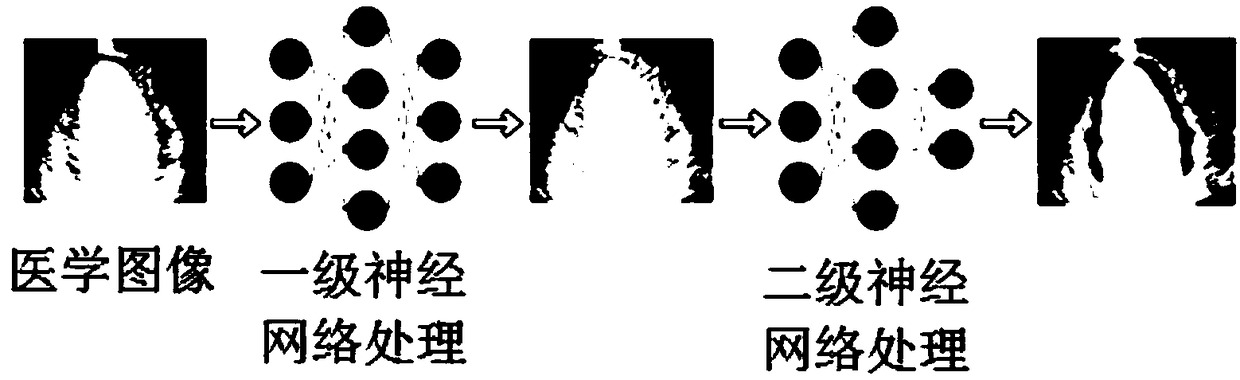
[0067] As mentioned above, taking myocardial perfusion contrast-enhanced cardiogram (MCE) as an example, 64 medical images of cases were randomly selected, and automatic myocardial contour delineation was realized according to the above steps. In this embodiment, the purpose of using cardiac ultrasound images is to accurately identify and outline the location of the myocardium, and the processed images are used for doctors to diagnose myocardial ischemia. It should be emphasized that, in some embodiments, the method of the invention is applicable to MRI, PET / CT, ultrasound, X-ray, photomicrograph, or other medical imaging. In addition, in some embodiments, various public medical image datasets can be used for testing, such as open source datasets of MedPix and the International Symposium on Biomedical Imaging (ISBI). The following table is the result of the present invention compared with other methods:

[0068] method

[0069] It can be seen from the above table th...

Embodiment 2

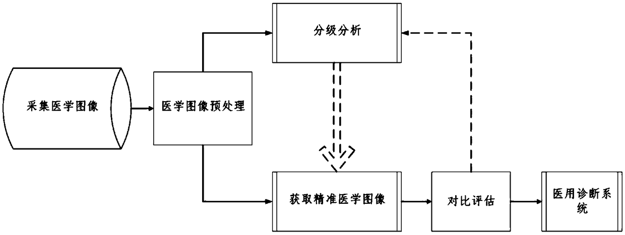
[0071] As mentioned above, a medical image diagnosis method includes the following steps:

[0072] ① Acquisition of medical images: Acquisition of relevant medical images from various medical equipment, including MRI, ultrasound, CT and X-ray;

[0073] ②Medical image preprocessing: perform format processing and medical image feature preprocessing on the medical images collected in step ①;

[0074] ③Image grading and delineation based on double convolutional neural network: the preprocessed medical image is delineated by double convolutional neural network, and the model parameters after grading analysis are stored in the computer, and the model parameters can be applied On a smaller data set, and achieve a good recognition effect;

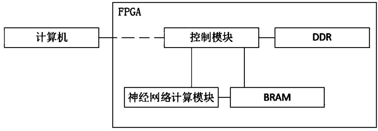
[0075] ④Hardware circuit design: According to the parameters of the secondary neural network model obtained in step ③, the hardware design of the neural network model on the FPGA. Because the first-level neural network only needs to divide the re...


PUM


Abstract

The present invention provides a medical image diagnostic method. The method comprises the steps of: collection of medical images, medical image preprocessing, classification analysis, obtaining of aprecious medical image and comparison assessment. A double convolutional neural network is employed to perform sketching of the medical images in batches, and the medical image diagnostic method can be widely applied to a small image data set to reduce the consumption of the calculation resources and obtain very high accuracy; a FPGA (Field Programmable Gate Array) is employed to achieve the wholedesign of a medical deagnostic tool, the power consumption of the FPGA is low to meet the energy conservation and emission reduction green economy development concept; the medical images with different types of diseases, different area crowds and different medical technical devices are employed to obtain corresponding parameters of a second-level neural network model so as to greatly facilitate doctors' usage and improve the diagnosis effect; and the economy cost of the neural network calculation unit based on the FPGA is low, so that medical image diagnostic method is suitable for each department of a general hospital and is conveniently applied to township-level medical institutions.

Description

technical field [0001] The invention relates to a medical image diagnosis method, which belongs to the cross technical field of artificial intelligence, medical image processing and customized calculation. Background technique [0002] Medical images are an important tool for diagnosing diseases. However, there is still a lack of reliable quantitative analysis tools for medical images at this stage, and most medical diagnoses still rely on doctors' experience and human judgment. Therefore, an accurate and reliable method and tool for automatic quantitative analysis of medical images is urgently needed. However, to achieve automated quantitative analysis, the first thing to achieve is the contouring of organs and tissues in medical images. In this regard, the current mainstream methods include traditional contour finding methods and machine learning methods. [0003] Traditional contour drawing methods include Active Contour Selection (ACS), discrete active contour, prior e...

Claims


Application Information

Patent Timeline
IPC IPC(8): G16H50/20G16H30/20
CPCG16H50/20G16H30/20
Inventor 李鹤庞雅薷
Owner 李鹤
Why Eureka
Try Eureka