

Method for determining biological activity of LAG3 (Lymphocyte Activation Gene-3) protein binding molecule
A molecular biology and molecular binding technology, applied in the field of measuring LAG3 protein binding molecular biological activity, which can solve the problems of heavy workload, poor repeatability, and inability to quantify specifically
- Summary
- Abstract
- Description
- Claims
- Application Information
AI Technical Summary
Problems solved by technology
Method used
Image

Examples
Embodiment 1
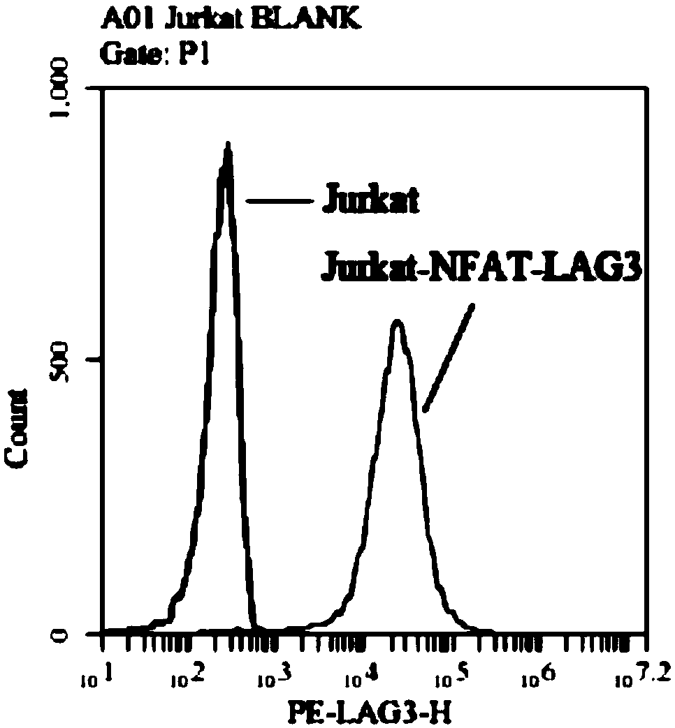
[0044] Embodiment 1, the screening of Jurkat-NFAT-LAG3 cell clone
[0045] 1. Protocells
[0046] The original Jurkat cells (Clone E6-1) involved in the present invention were purchased from the Shanghai Cell Bank of the Chinese Academy of Sciences and subcultured and preserved in our laboratory. The cell viability rate used by the cells is over 95%.
[0047] Jurkat cell clones were resuspended with OPTI-MEM (GIBCO, 31985062) medium, transferred to an electric shock cup provided with an electroporation instrument (Bio-Rad, Gene Pulser Xcell), the plasmid was added and mixed gently, and then electroshocked, and then all the cells were suspended The solution was transferred to a complete medium (90% 1640+10% FBS) in a clean six-well plate (Costar, 3516), and cultured statically in a 37°C incubator (Thermo, BB150I) for 24 hours.
[0048] 2. Plasmid
[0049] The plasmid pGL4.30[luc2P / NFAT-RE / Hygro] (Promega, E848A) with NFAT reporter element, and the pIRES-puro3 plasmid (Clonte...
Embodiment 2
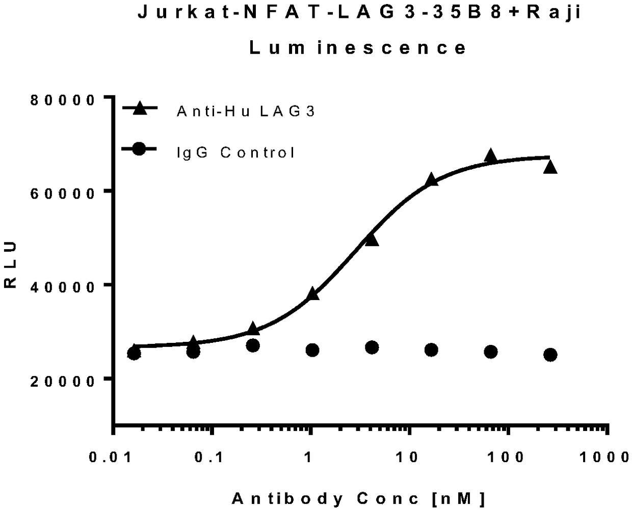
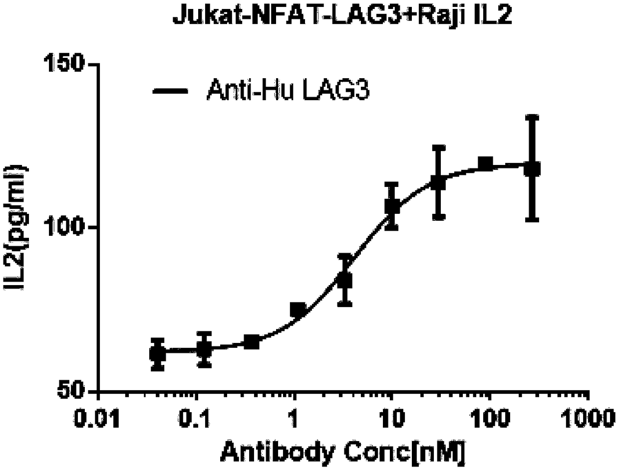
[0061] Embodiment 2, reporter gene experimental method
[0063] The cell clone involved in the present invention is the Jurkat-NFAT-LAG3 cell clone after transfection screening and identification in our laboratory, and the clone number is 35B8. It was verified as a cell clone with high expression of NFAT reporter element and LAG3 protein. The Raji cells involved in the present invention are human Burkitt's lymphoma cells, purchased from the Shanghai Cell Bank of the Chinese Academy of Sciences, and subcultured and preserved in our laboratory. After the cells were taken out of the liquid nitrogen tank, they were immediately put into a 37°C water bath to thaw until a small piece of ice remained, diluted with complete medium (90% 1640+10% FBS) and transferred to a clean 15mL centrifuge tube (Corning, 430791). Centrifuge at 400×g to discard the supernatant, resuspend the cells with an appropriate volume of complete medium and inoculate the cells on a ce...
Embodiment 4
[0085] Embodiment 4, reporter gene experiment optimization
[0086] 1. The ratio relationship between Jurkat-NFAT-LAG3 and Raji cells
[0087] The cells involved in the present invention are Jurkat-NFAT-LAG3 cell clones and Raji cells. When cell sampling, the cell number of Jurkat-NFAT-LAG3 was fixed at 1×10 5 One / well, Raji cells were sampled as 1 / 4, 1 / 6 and 1 / 8 of the number of Jurkat-NFAT-LAG3 cells, respectively. Then the reporter gene experiment was carried out under the conditions of fixed SED concentration of 100ng / mL and 150ng / mL, and the cell number matching conditions with good luminescence intensity and signal window were selected. The effect of different cell ratio conditions on the reporter gene results is as follows: Figure 4 As shown, under the two SED concentration conditions, different cell ratios can show that the intensity of the luminescent signal increases with the increase of the antibody concentration, and the cell ratios of 4:1 and 8:1 are better. ...
PUM
 Abstract
Description
Claims
Application Information