An intima-media segmentation method for dual-channel intravascular ultrasound images

An ultrasound imaging, dual-channel technology, applied in the field of medical image processing, can solve the problems of the decrease in the segmentation accuracy of the medial intima region, the inability to meet the complex situation of intravascular ultrasound images, and the inability of the network to find the optimal solution, and to increase the amount of information. , the effect of suppressing ultrasonic speckle and improving segmentation efficiency

Active Publication Date: 2018-12-14
SOUTH CHINA UNIV OF TECH
View PDF3 Cites 3 Cited by
  • Summary
  • Abstract
  • Description
  • Claims
  • Application Information

AI Technical Summary

Problems solved by technology

However, this method is easily affected by speckles, noise and artifacts in the ultrasound image, making the network unable to find its optimal solution, which eventually leads to a decrease in the accuracy of the media-intima region segmentation.
Unable to meet the complex situation of clinical intravascular ultrasound imaging

Method used


Image

  • An intima-media segmentation method for dual-channel intravascular ultrasound images
  • An intima-media segmentation method for dual-channel intravascular ultrasound images
  • An intima-media segmentation method for dual-channel intravascular ultrasound images

Examples


Embodiment 1

[0045] The method for segmenting the intima of a blood vessel in a dual-channel intravascular ultrasound image provided in this embodiment includes the following steps:

[0046] Step 1: A total of 753 intravascular ultrasound images are collected, 600 of which are used as training set data, and 153 are used as independent test set; the image size is 512×512.

[0047] Step 2: The anisotropic diffusion filtering process is as follows:

[0048] For a grayscale image I(x,y) of size 512×512. According to the iteration formula as follows:

[0049]

[0050] where I t Represents the current pixel value of the image, t is 300, and λ is 0.1, both of which can control the smoothness of the image. The constant term k in the four-neighborhood diffusion coefficient is 15.

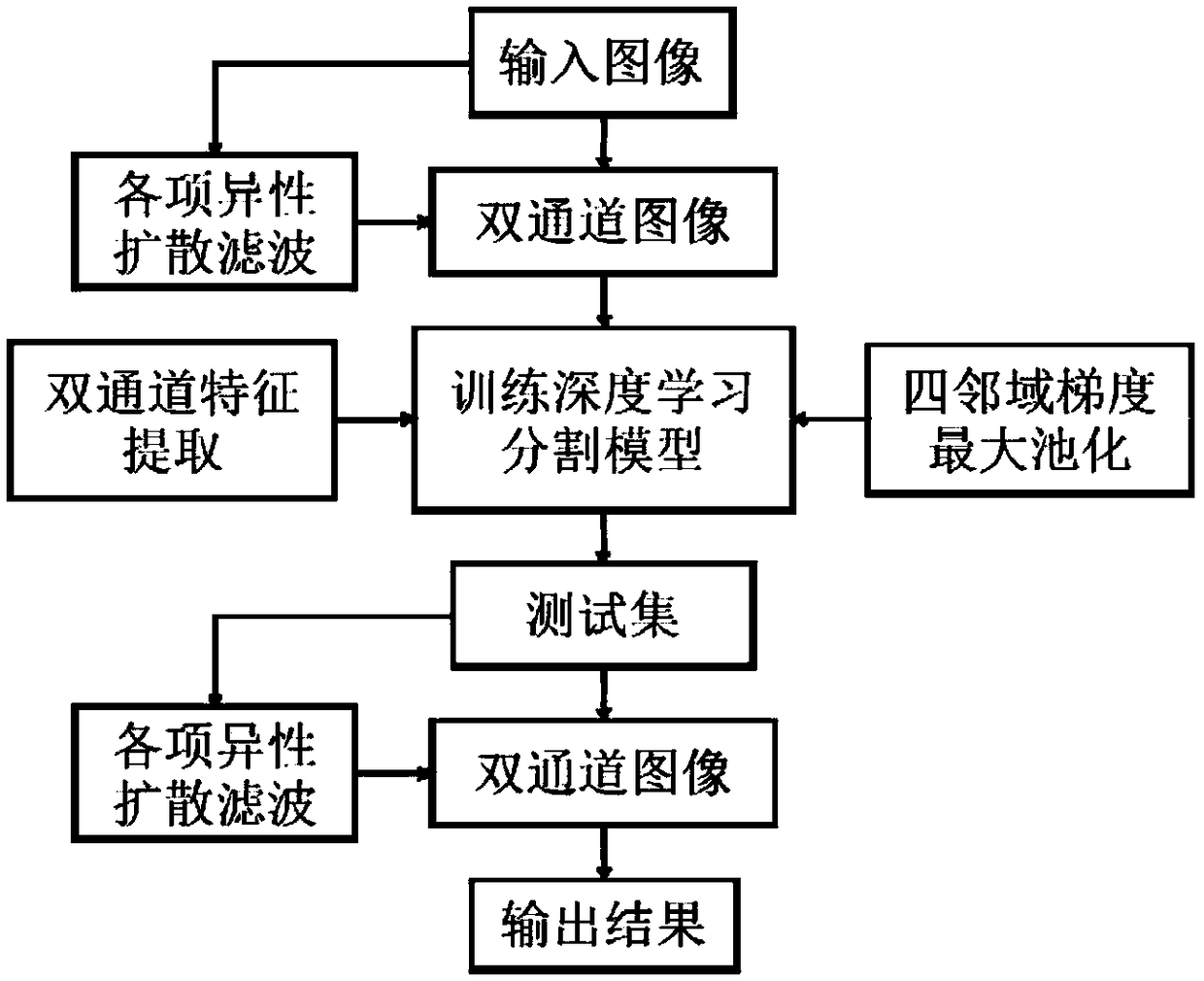
[0051] Step 3: As attached figure 1 In the flow chart shown, the dual-channel image data construction process is as follows:

[0052]After the original image A of 512×512 size is processed by anisotropic diffusi...

Embodiment 2

[0060] The method for segmenting the intima of a blood vessel in a dual-channel intravascular ultrasound image provided in this embodiment includes the following steps:

[0061] Step 1: Carry out polar coordinate transformation on the collected images, and transform the 512×512 images into 384×384 images.

[0062] Step 2: The anisotropic diffusion filtering process is as follows:

[0063] For a grayscale image I(x,y) of size 384×384. According to the iteration formula as follows:

[0064]

[0065] where I t Represents the current pixel value of the image, t is 100, and λ is 0.15. The constant term k in the four-neighborhood diffusion coefficient is 15.

[0066] Step 3: Construct dual-channel image data.

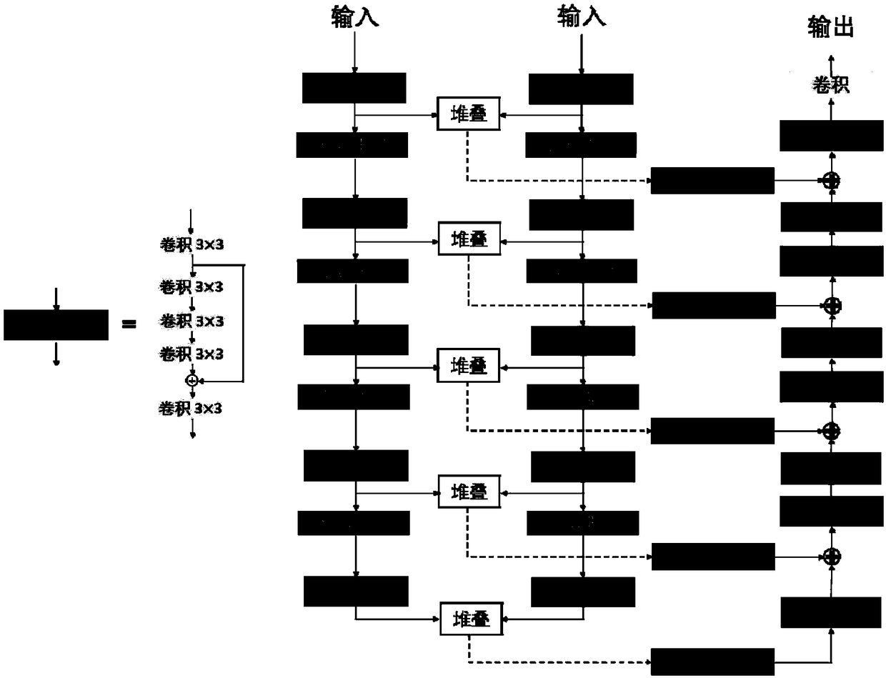
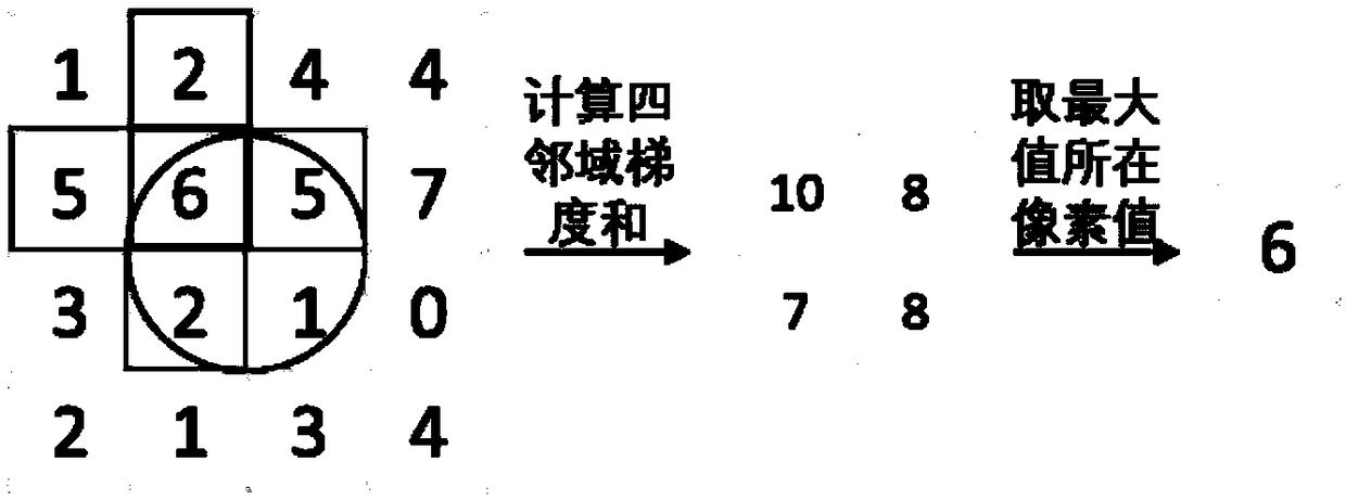
[0067] Step 4: Train the network parameters with the input image. The pooling layer uses neighborhood gradient max pooling.

[0068] Step Five: Attach Figure 5 Comparison of model test results showing polar imagery and manually delineated standards.


PUM


Abstract

The invention discloses an intima-media segmentation method for dual-channel intravascular ultrasound images, which comprises the following steps: 1) collecting blood vessel intima-media ultrasound images, artificially delineating intima-media boundaries, and establishing training and testing sample sets; 2) carrying out anisotropic diffusion filtering processing on the images of the training sample set; 3) superimposing the filtered new images and the original images to form a dual-channel image; 4) establishing a depth learning segmentation model, pooling the maximum neighborhood gradient, and inputting a training set for training; 5) carrying out anisotropic diffusion filtering on the image of the testing set, composing the filtered image and the original image into a dual-channel image, inputting the trained depth learn segmentation model, and segmenting to obtain the segmented blood vessel intima boundary. The method of the invention can accurately and effectively extract the blood vessel intima region.

Description

technical field [0001] The invention relates to the technical field of medical image processing, in particular to a method for segmenting the intima of a blood vessel for a dual-channel intravascular ultrasonic image. Background technique [0002] In recent years, the global morbidity and mortality of coronary heart disease are on the rise. Its pathogenesis is coronary atherosclerotic lesions, leading to stenosis or even occlusion of the vascular lumen. At present, intravascular ultrasound (Intravascular Ultrasound, IVUS) can be used clinically to obtain real-time intravascular cross-sectional images, which can be used to analyze the thickness of the blood vessel wall and the tissue composition of atherosclerotic plaques, helping doctors to judge the condition, determine the lesion site, and design a treatment plan . [0003] In IVUS images, doctors pay most attention to the intima-media region of blood vessels, so the use of computer-aided diagnosis to realize the automat...

Claims


Application Information

Patent Timeline
Patent Type & Authority Applications(China)
IPC IPC(8): G06T7/11G06N3/04
CPCG06T7/11G06T2207/30101G06T2207/20096G06N3/045
Inventor 郭圣文吴宇鹏乔垣方任力梁稳生黄美萍
Owner SOUTH CHINA UNIV OF TECH
Why Eureka
Try Eureka