Three dimensional (3D) printed PCL-PDA-AgNPs scaffold for anti-infective bone tissue engineering as well as preparation method and application of 3D printed PCL-PDA-AgNPs scaffold

A technology of bone tissue engineering and 3D printing, applied in tissue regeneration, additive processing, medical science, etc., can solve the problems of high price, immune rejection of bioactive factors, low processing performance, etc., and improve hydrophilicity and biophase Capacitance, promotion of new bone tissue formation, beneficial effect of exchange

Active Publication Date: 2018-12-21
南京冬尚生物科技有限公司
View PDF4 Cites 6 Cited by
  • Summary
  • Abstract
  • Description
  • Claims
  • Application Information

AI Technical Summary

Problems solved by technology

However, these materials all have certain defects, including: 1. HA, CPC and other materials have high brittleness and low processing performance, and it is difficult to process and plasticize a specific three-dimensional shape, which often depends on the three-dimensional configuration of the material itself; 2. PGA, Materials such as PLA have low biological activity, elastic modulus, tensile strength, and three-dimensional finishing methods; Medication characteristics in the infection

Method used


Image

  • Three dimensional (3D) printed PCL-PDA-AgNPs scaffold for anti-infective bone tissue engineering as well as preparation method and application of 3D printed PCL-PDA-AgNPs scaffold
  • Three dimensional (3D) printed PCL-PDA-AgNPs scaffold for anti-infective bone tissue engineering as well as preparation method and application of 3D printed PCL-PDA-AgNPs scaffold
  • Three dimensional (3D) printed PCL-PDA-AgNPs scaffold for anti-infective bone tissue engineering as well as preparation method and application of 3D printed PCL-PDA-AgNPs scaffold

Examples


Embodiment 1

[0043] A 3D printing PCL-PDA-AgNPs anti-infection bone tissue engineering scaffold, which is prepared by the following method:

[0044] Step 1: Prepare 3D printed PCL scaffold

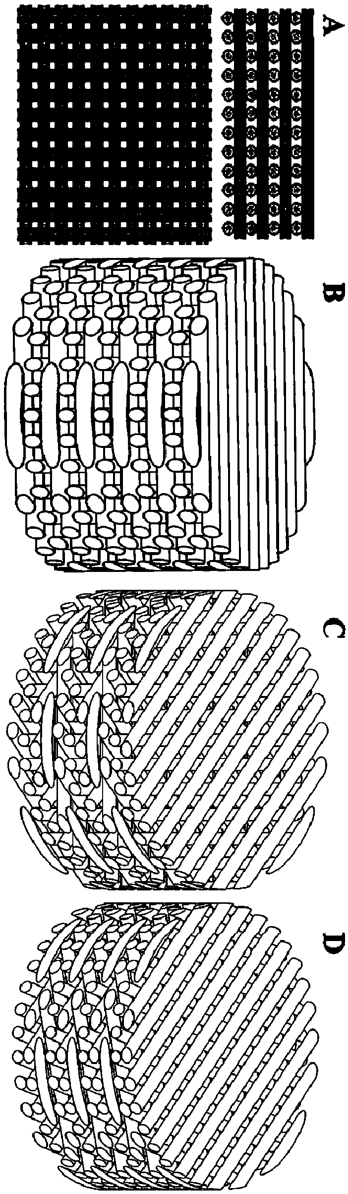
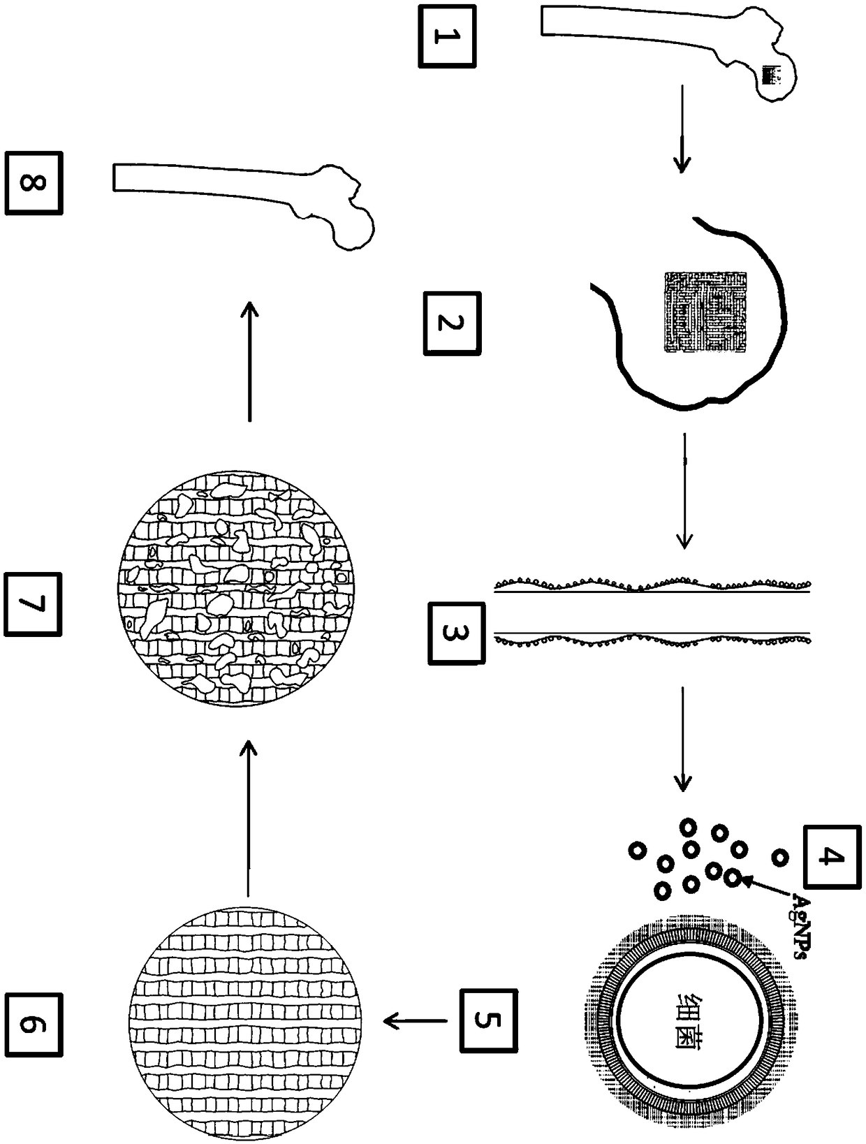
[0045] Such as figure 1 As shown in (A), the PCL is heated to a liquid state through the heating system of the melt-extruded 3D printing, extruding the molded fiber bundles, and obtaining the 3D scaffold PCL by superimposing and arranging different layers of the fiber bundles;

[0046] Step 2: Prepare 3D printed PCL-PDA bracket

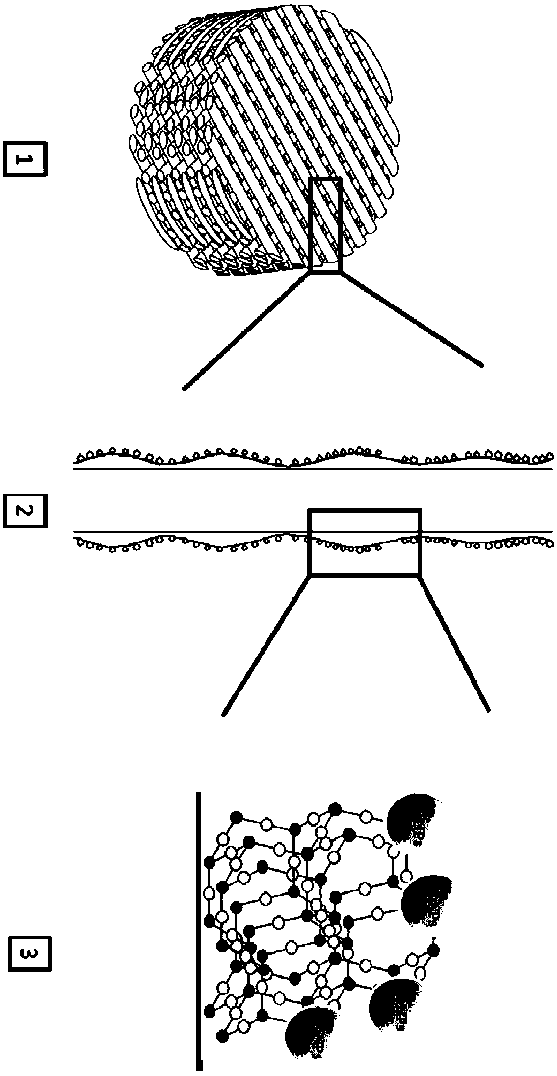
[0047] Dissolve 0.61g Tris in 500mL water, then add HCL to adjust the pH of the system to 8-9, and prepare a Tris-HCL solution; dissolve 1g dopamine in the Tris-HCL solution, stir to dissolve, and configure it as a DA-Tris-HCL solution ;Add the 3D scaffold PCL to the DA-Tris-HCl solution, stir under the condition of avoiding light, and take out the scaffold after the DA self-polymerizes on the surface of the PCL to form PDA, and obtain the 3D printed PCL-PDA scaffold;

[00...

Embodiment 2

[0051] A 3D printing PCL-PDA-AgNPs anti-infection bone tissue engineering scaffold, which is prepared by the following method:

[0052] Step 1: Prepare 3D printed PCL scaffold

[0053] Such as figure 1 As shown in (B), the PCL is heated to a liquid state through the heating system of the melt-extruded 3D printing, and the molded fiber bundle is extruded, and the 0-90° stacked arrangement of the fibers is a square-pore 3D support PCL;

[0054] Step 2: Prepare 3D printed PCL-PDA bracket

[0055] Dissolve 0.4g Tris in 500mL water, then add HCL to adjust the pH value of the system to 8-9, and prepare a Tris-HCL solution; dissolve 1g dopamine in the Tris-HCL solution, stir to dissolve, and configure it as a DA-Tris-HCL solution ;Add the 3D scaffold PCL to the DA-Tris-HCl solution, stir under the condition of avoiding light, and take out the scaffold after the DA self-polymerizes on the surface of the PCL to form PDA, and obtain the 3D printed PCL-PDA scaffold;

[0056] Step 3: P...

Embodiment 3

[0059] A 3D printing PCL-PDA-AgNPs anti-infection bone tissue engineering scaffold, which is prepared by the following method:

[0060] Step 1: Prepare 3D printed PCL scaffold

[0061] Such as figure 1 As shown in (C), the PCL is heated to a liquid state through the heating system of the melt-extruded 3D printing, and the molded fiber bundles are extruded, and the fiber bundles are stacked at 0-60-120-180° to obtain a 3D scaffold PCL;

[0062] Step 2: Prepare 3D printed PCL-PDA bracket

[0063] Dissolve 0.8g Tris in 500ml of water, then add HCL to adjust the pH of the system to 8-9, and prepare a Tris-HCL solution; dissolve 1g of dopamine in the Tris-HCL solution, stir to dissolve, and configure it as a DA-Tris-HCL solution ;Add the 3D scaffold PCL to the DA-Tris-HCl solution, stir under the condition of avoiding light, and take out the scaffold after the DA self-polymerizes on the surface of the PCL to form PDA, and obtain the 3D printed PCL-PDA scaffold;

[0064] Step 3: ...


PUM


Abstract

The invention relates to a three dimensional (3D) printed PCL-PDA-AgNPs scaffold for anti-infective bone tissue engineering as well as a preparation method and application of the 3D printed PCL-PDA-AgNPs scaffold, belonging to bioengineered human implants. According to the method, poly(epsilon caprolactone) is used as a base material, polydopamine is used as a surface modification material, and silver nanoparticles with anti-infective biological properties are combined for use, so that the biological functions of anti-infection, bone growth promotion and bone defect repair are realized. The 3Dprinted PCL-PDA-AgNPs scaffold for the anti-infective bone tissue engineering has the advantages of being simple and reliable in structure, controllable in appearance and micro-structure, reliable inmechanical properties, controllable in ion release property, convenient in implantation, less in trauma and low in cost.

Description

technical field [0001] The invention relates to 3D printing PCL-PDA-AgNPs anti-infection bone tissue engineering scaffold and its preparation method and application, which belongs to the bioengineered human body implant and can be used to treat various pathogenic bacteria, such as: Staphylococcus aureus, streptococcus suppurativa Treatment of acute and chronic osteomyelitis, septic arthritis bone destruction caused by coccus, β-hemolytic streptococcus, Bacillus anthracis, diphtheria bacillus, etc., and repair of bone defects after infection. Background technique [0002] Infection is defined as a disruption of the balance between the host tissue and the local concentration of microorganisms, generally considered to be greater than 10 5 Each gram of tissue or the presence of pathogenic bacteria such as Staphylococcus aureus, Streptococcus pyogenes, and β-hemolytic streptococcus is considered to be an infection. Among many infectious diseases, bone and joint infection is not ...

Claims


Application Information

Patent Timeline
IPC IPC(8): A61L27/56A61L27/18A61L27/02A61L27/54A61L27/50A61L27/58B33Y10/00B33Y80/00
CPCA61L27/025A61L27/18A61L27/50A61L27/54A61L27/56A61L27/58A61L2300/104A61L2300/404A61L2430/02B33Y10/00B33Y80/00C08L67/04C08L79/02
Inventor 王黎明姚庆强周进
Owner 南京冬尚生物科技有限公司
Why Eureka
Try Eureka