Humanized nanometer antibody resistant to PD-L1 and application thereof

A PD-L1, nanobody technology, applied in the field of biomedicine or biopharmaceuticals, can solve problems such as reduced T cell immune effect

Active Publication Date: 2018-12-28
HUALAN BIOLOGICAL ENG INC +1
View PDF2 Cites 15 Cited by
  • Summary
  • Abstract
  • Description
  • Claims
  • Application Information

AI Technical Summary

Problems solved by technology

However, activation of the PD-1 / PD-L1 signaling pathway changes the local microenvironment of the tumor and reduces the immune effect of T cells, thereby mediating tumor immune escape and promoting tumor growth

Method used


Image

  • Humanized nanometer antibody resistant to PD-L1 and application thereof
  • Humanized nanometer antibody resistant to PD-L1 and application thereof
  • Humanized nanometer antibody resistant to PD-L1 and application thereof

Examples


Embodiment 1

[0166] Example 1: Expression and purification of human PD-L1 protein

[0167] (1) The nucleotide sequence of human PD-L1 was synthesized on the pCDNA3.1(-) vector, and then its extracellular segment sequence was subcloned into the pFMSE-IgG1 vector;

[0168] (2) Extract the constructed pFMSE-IgG1-hPD-L1 (ECD) plasmid with the Omega plasmid extraction kit;

[0169] (3) Culture HEK293F cells to an OD of 2.0×10 6 a / mL;

[0170] (4) Mix the plasmid and the transfection reagent PEI 1:3 evenly, let it stand for 10 minutes, and then add it to HEK293F cells at 37°C, 6% CO 2 Cultivate in a shaker incubator for 5-6 days;

[0171] (5) Cell supernatant was collected and combined with Protein A beads for 1 h at room temperature;

[0172] (6) After washing the beads with phosphate buffer solution pH 7.0, the protein was eluted with 0.1M pH 3.0 Glycine;

[0173] (7) Ultrafilter the eluted protein into PBS, measure the yield and take a sample for SDS-PAGE to test the purity of the antigen,...

Embodiment 2

[0175] Example 2: Construction of PD-L1 Nanobody Library

[0176] (1) Mix 1 mg of hPD-L1(ECD)-Fc antigen with Freund's adjuvant in equal volume, immunize a Xinjiang Bactrian camel once a week, and immunize 3 times in total to stimulate B cells to express antigen-specific nanobodies ;

[0177] (2) After the 3 times of immunization, extract 100mL camel peripheral blood lymphocytes and extract total RNA;

[0178] (3) Synthesize cDNA and amplify VHH by nested PCR;

[0179] (4) Digest 20 μg of pMECS phage display vector (supplied by Biovector) and 10 μg of VHH with restriction enzymes Pst I and Not I and connect the two fragments;

[0180] (5) The ligation product was transformed into electroporation-competent cells TG1, and the PD-L1 nanobody library was constructed and the storage capacity was determined. The storage capacity was 1.0×10 9 CFU;

[0181] At the same time, 24 clones were randomly selected for colony PCR detection, and the results showed that the insertion rate o...

Embodiment 3

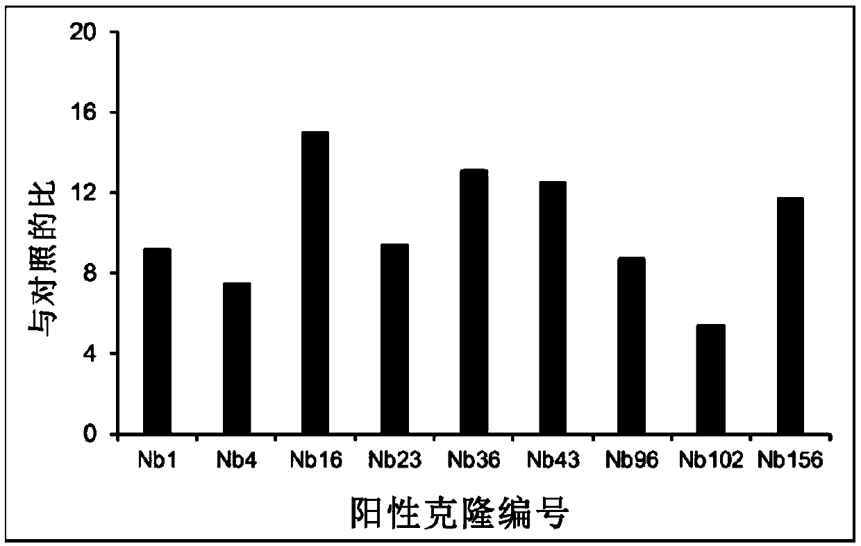
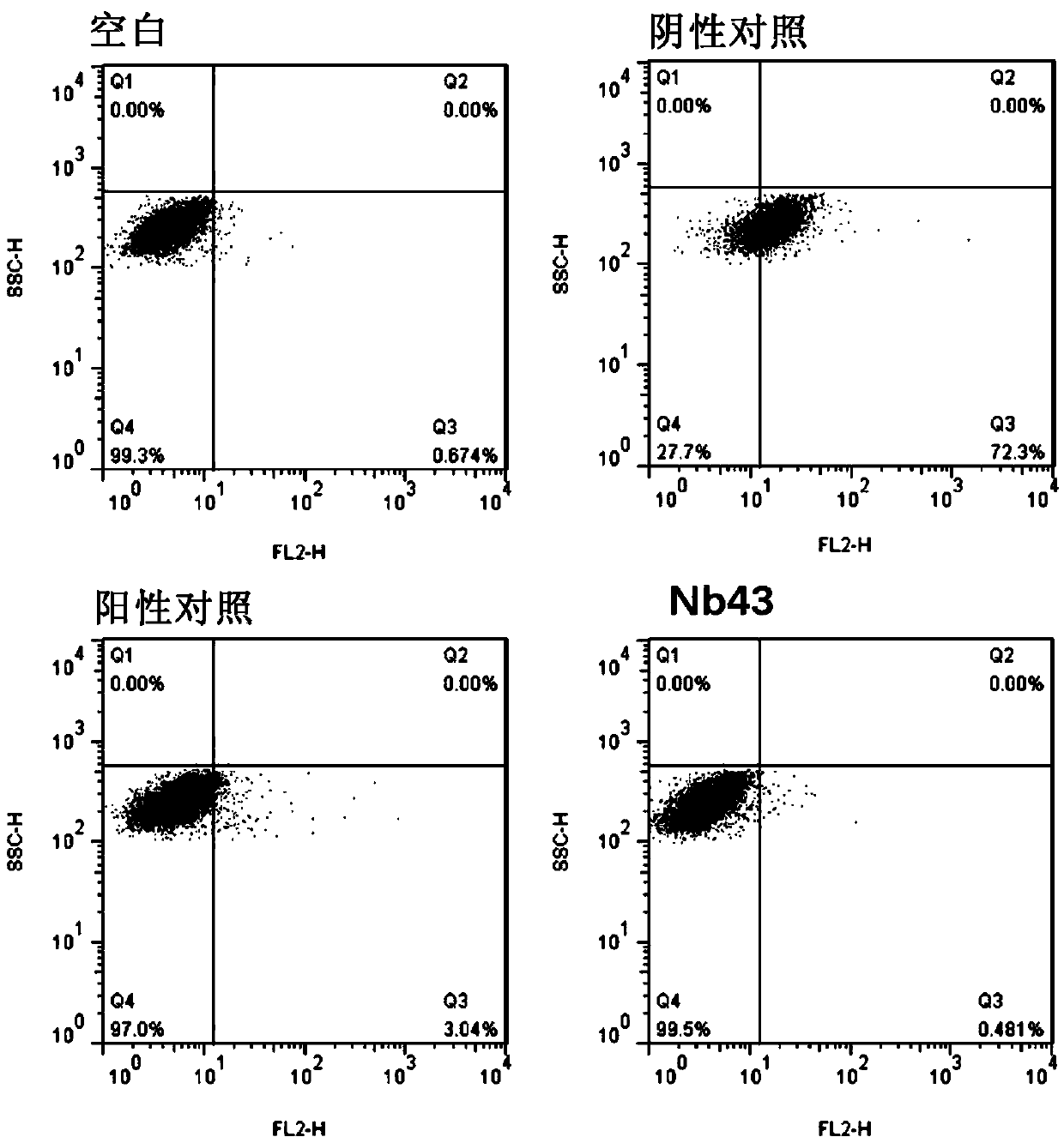
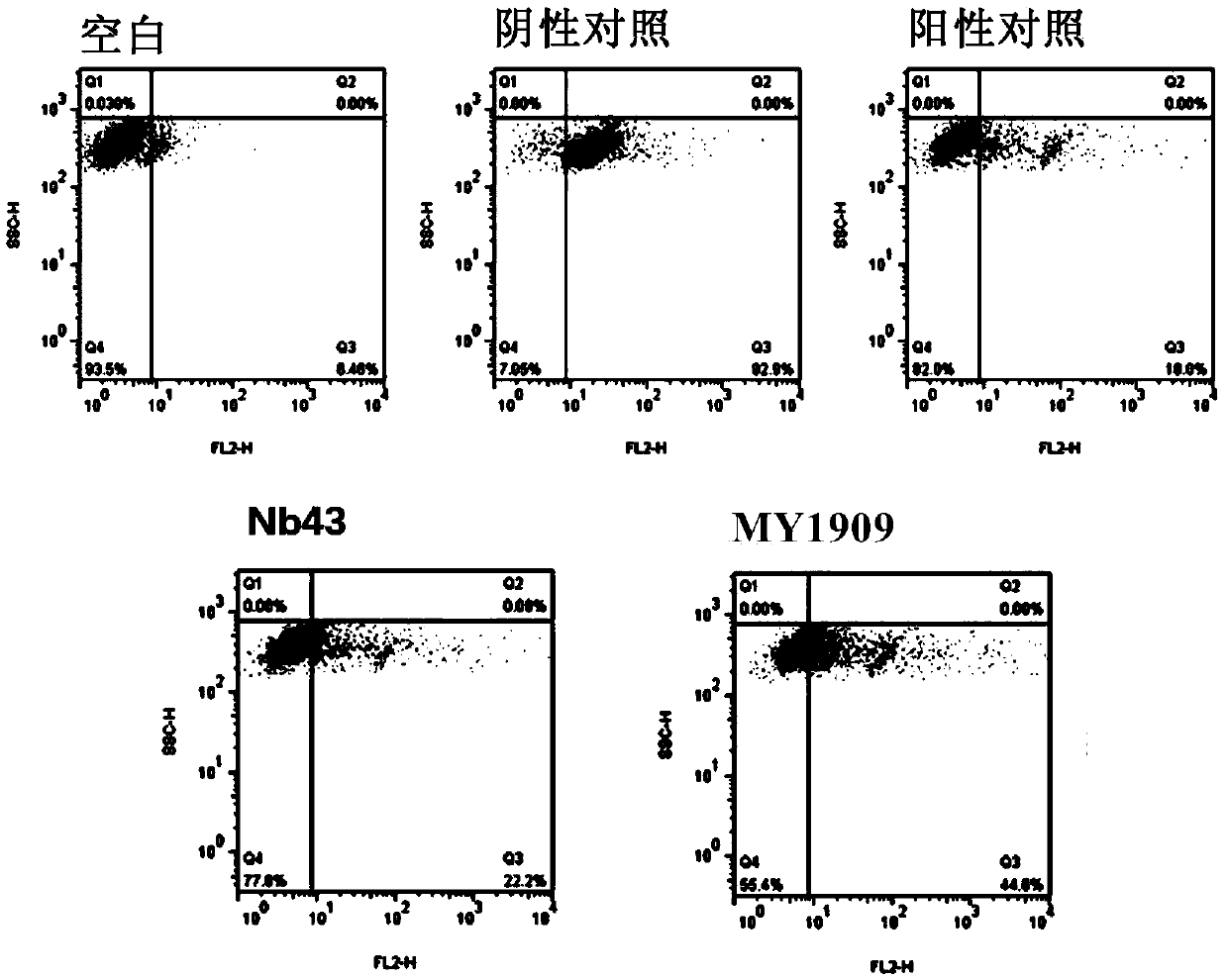
[0182] Example 3: Screening and Identification of PD-L1 Nanobodies

[0183] Antibody Screening:

[0184] (1) Dissolve in 100mM NaHCO 3 , 10 μg hPD-L1(ECD)-Fc antigen (10 μg Fc inNaHCO 3 As a control) was coupled on a NUNC microtiter plate and placed overnight at 4°C;

[0185] (2) Add 100 μL 0.01% BSA the next day, and block at room temperature for 0.5 h;

[0186] (3) After 2h, add 100μL phage (2×10 11 CFU immunized camel nanobody phage display gene library), at room temperature for 0.5h;

[0187] (4) Wash 5 times with 0.05% PBS+Tween-20 to wash off non-specific phage;

[0188] (5) Dissociate the phage that specifically binds to PD-L1 with 100mM triethanolamine, infect Escherichia coli TG1 cells in the logarithmic phase, and culture at 37°C for 1 hour to generate and purify the phage for the next round of screening , the same screening process was repeated for 3 rounds, and the enrichment factors were no enrichment, 11.4 times and 270 times respectively.

[0189] Use pha...


PUM


Abstract

The invention provides a humanized nanometer antibody resistant to PD-L1 and application thereof and particularly provides a nanometer antibody resistant to a protein programmed-death ligand (PD-L1).The provided nanometer antibody resistant to PD-L1 can effectively interdict the combination between the PD-L1 and programmed death molecules 1 (PD-1), and therefore an inhibiting effect of the PD-L1on T cells capable of expressing the PD-1 can be successfully counteracted. The PD-L1 nanometer antibody can be applied to prevention, diagnoses and treatment of diseases correlated with the PD-L1. The invention also provides a humanized PD-L1 nanometer antibody sequence, and the humanized nanometer antibody still has the function of interdicting the combination between the PD-L1 and the PD-1, ishigh in affinity and specificity and can serve as a tumor immunity target antibody drug.

Description

technical field [0001] The present invention relates to the technical field of biomedicine or biopharmaceuticals, and more specifically relates to anti-PD-L1 nanobody and its application. Background technique [0002] Cell-mediated cellular immune response is the basis of anti-tumor immunity, and the activation and proliferation of T cells require dual signals. That is, in addition to the first signal generated by the combination of T cell receptor (TCR) and the MHC-antigen peptide complex expressed on the surface of antigen-presenting cells (APCs); it also requires a variety of co-stimulatory molecules to participate in the second signal provided ( co-stimulatory signal). [0003] In the initial stage of the immune response, under the joint action of the first signal provided by TCR and the co-stimulatory signal, T cells reach their activation threshold and are activated, producing effector and memory cells, and exerting immune defense functions. A series of co-stimulate ...

Claims


Application Information

Patent Timeline
Patent Type & Authority Applications(China)
IPC IPC(8): C07K16/28C12N15/13G01N33/68A61K39/395A61P35/00A61P35/02
CPCA61P35/00A61P35/02C07K16/2827C07K2317/76C07K2317/569C07K2317/565C07K2317/24A61K39/395A61K51/10C07K16/28G01N33/68
Inventor 安康安文琪范蓓马小伟潘若文张宝献
Owner HUALAN BIOLOGICAL ENG INC
Why Eureka
Try Eureka