
Fluorescence in situ hybridization probe for detecting KIAA1549-BRAF fusion gene and preparation method and application thereof

A KIAA1549-BRAF, fluorescence in situ hybridization technology, applied in the field of molecular biology, can solve the problems of incomplete distinction between positive and negative samples, inability to distinguish break signal points, short distance, etc., and achieve high signal-to-noise ratio and short hybridization time Short, wide selection of effects

Active Publication Date: 2019-01-18
WUHAN HEALTHCHART BIOLOGICAL TECH
View PDF3 Cites 1 Cited by
  • Summary
  • Abstract
  • Description
  • Claims
  • Application Information

AI Technical Summary

Problems solved by technology

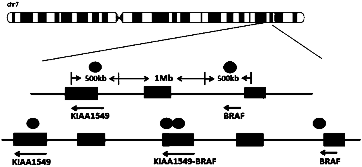
Since the design region of the existing BRAF breakage probe is 550Kb around the BRAF gene breakage area, when the BRAF gene breaks, the distance between the signal points is 1.4Mb, which is relatively short and cannot be distinguished from the breakage signal points; and KIAA1549 / BRAF fusion gene probe design region ( figure 2 ) fusion regions of the two genes respectively, and one more fusion signal appeared in the middle of the two signal points after the fusion, but the distance between the three signal points was about 1.0Mb, and the break signal points could not be distinguished as well.
Therefore, neither the conventional BRAF breakage probe nor the KIAA1549 / BRAF fusion gene probe can fully distinguish positive and negative samples at present

Method used


Image

  • Fluorescence in situ hybridization probe for detecting KIAA1549-BRAF fusion gene and preparation method and application thereof
  • Fluorescence in situ hybridization probe for detecting KIAA1549-BRAF fusion gene and preparation method and application thereof
  • Fluorescence in situ hybridization probe for detecting KIAA1549-BRAF fusion gene and preparation method and application thereof

Examples


Embodiment 1

[0045] Example 1 Preparation of KIAA1549-BRAF fusion gene rapid detection probe

[0046] The preparation of the KIAA1549-BRAF fusion gene rapid detection probe of the present invention includes the following steps:

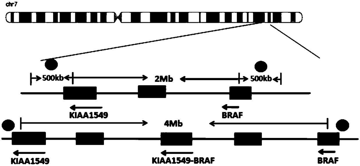
[0047] (1) Download from UCSC Genome Browser the genomic non-repetitive sequence of the region covered by the KIAA1549 and BRAF gene probes. The KIAA1549 probe covers the region: chr7 138,361,140-138,861,139 (starting position is the direction of exon 16 of KIAA1549 gene toward the centromere 500Kb region); BRAF gene probe coverage area is: chr7 140,787,547-141,287,546 (starting position is the 500kb region from exon 9 of BRAF gene to the telomere direction); the obtained sequence is saved in fasta format, and the repetitive sequence region in the genome is replaced with N;

[0048] (2) Use the perl plug-in program chunks.pl to divide the genomic non-repetitive sequence in the area covered by the KIAA1549 gene probe obtained in step (1) into 1kb blocks; import the divid...

Embodiment 2

[0055] Example 2 Method of using KIAA1549-BRAF fusion gene rapid detection probe

[0056] The method for using the KIAA1549-BRAF fusion gene rapid detection probe prepared in Example 1 to detect the KIAA1549-BRAF fusion gene specifically includes the following steps:

[0057] (1) Sample processing: Take the paraffin section sample with a thickness of 4 to 5 microns that has been baked, and immerse it in preheated 65°C dewaxing agent I for 10 to 15 minutes; take out the slide and immerse it in preheated 65°C dewaxing Reagent II for 10-15 minutes; after taking out the slide, immerse it in 100% ethanol, 85% ethanol, and 70% ethanol for 2 to 3 minutes at room temperature; take out the slide and immerse it in preheated 65℃ deionized water for 3~ 5 minutes; take out the slide and soak in 95℃ deionized water for 30-40 minutes, pre-cool the slide in cold water for 1 min; take out the slide and immerse it in preheated 37℃ protease working solution for 20-40 minutes; take out the slide and s...


PUM


Abstract

The invention belongs to the field of molecular biology, in particular to a fluorescence in situ hybridization probe for detecting KIAA1549-BRAF fusion gene and preparation method and application thereof. The fluorescence in situ hybridization probe comprises two probe libraries, a KIAA1549 gene hybridization probe library: the starting position of the probe is designed in the range of 200 Kb around the KIAA1549 gene breakpoint, the probe extends toward the direction of the centromere, and the length of the probe is 300 to 1000 Kb; BRAF gene hybridization probe library: The starting position of the probe is designed in the range of 200 Kb around the breakpoint of BRAF gene, and the probe extends to the telomere. The length of the probe is 300 - 1000 Kb. The invention designs probes in twofusion gene outer regions respectively, realizes the detection of two fusion genes which are relatively close on the same chromosome, and can directly reflect the fusion state of the two genes on thecell level.

Description

Technical field [0001] The invention belongs to the field of molecular biology, and specifically relates to a fluorescent in situ hybridization probe for detecting the KIAA1549-BRAF fusion gene, and a preparation method and application thereof. Background technique [0002] Pilocytic astrocytoma (PA) is a primary intracranial tumor graded by WHO. It is the most common glioma in children's neurosurgery, accounting for about 25% of brain tumors. Both KIAA1549 gene and BRAF gene are located in the q34 region of chromosome 7. The tandem duplication of BRAF gene leads to the fusion of KIAA1549 / BRAF gene, which is more common in pilocytic astrocytoma (60%-80%). According to the different fusion positions of KIAA1549 and BRAF genes, 5 different fusion variants have been reported recently, namely: KIAA1549ex16-BRAF ex9, KIAA1549ex15-BRAF ex9, andKIAA1549ex16-BRAF ex11, KIAA1549ex18-BRAF ex10 and KIAA1549ex19-BRAF ex9; Three types accounted for 45%, 28% and 5% of the total variation in s...

Claims


Application Information

Patent Timeline
IPC IPC(8): C12Q1/6886C12Q1/6841C12N15/11
CPCC12Q1/6841C12Q1/6886C12Q2563/107
Inventor 吕玉琦李先洋李倩李雪梅叶伦程弘夏陈刚
Owner WUHAN HEALTHCHART BIOLOGICAL TECH
Learn More