System for bright field image simulation

An image and bright field technology, applied in the field of fluorescence image processing, can solve problems such as wrong diagnosis, wrong treatment advice, wrong immune cells as cancer cells, etc.

Active Publication Date: 2019-02-05
F HOFFMANN LA ROCHE & CO AG
View PDF10 Cites 8 Cited by
  • Summary
  • Abstract
  • Description
  • Claims
  • Application Information

AI Technical Summary

Problems solved by technology

Alternatively, the laboratory may simply have changed the staining protocol without notifying the pathologist of the change
As a result, the pathologist may mistake immune cells for cancer cells and may generate wrong diagnoses and wrong treatment recommendations

Method used


Image

  • System for bright field image simulation
  • System for bright field image simulation
  • System for bright field image simulation

Examples


Embodiment Construction

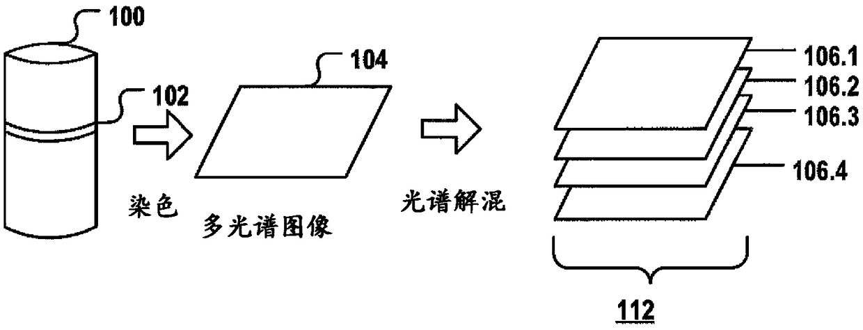
[0148] figure 1 Depicted is the acquisition of multiple monochrome images from a multispectral image via color deconvolution.

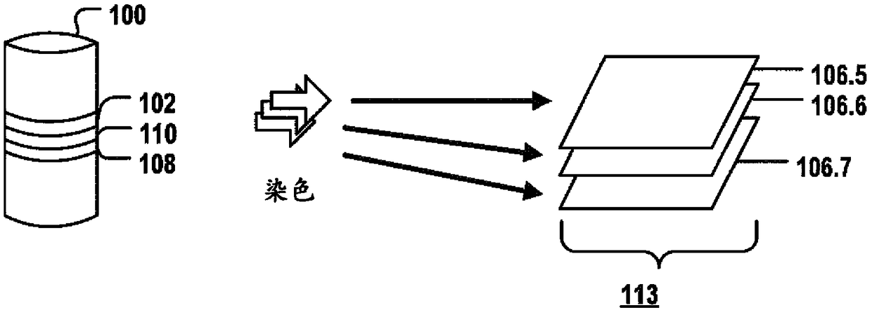
[0149] Tissue samples are provided first. For example, one or more biopsy samples are taken as part of the diagnosis of many cancer types, such as colorectal cancer. The biopsy sample is cut into one or more thin tissue layers 102 . layer 102 can be stained with one or more fluorescent stains that selectively stain specific biomarkers, cells, and / or organelles, and a multispectral image 104 is acquired from layer 102 to capture meaningful biomedical features, It may allow tumors to be classified and / or may allow prediction of clinical outcome and / or generation of treatment recommendations. An image acquisition system, such as a slide scanner or microscope, can acquire a multispectral image 114, which can contain spectral information for multiple different fluorescent stains, and can contain autofluorescent signals from the tissue layer 102 on the s...


PUM


Abstract

The invention relates to an image processing system for generating a simulated digital bright field IHC or ISH image from monochromatic, fluorescence images (106.1-106.7) of a tissue sample (100). Thetissue sample comprises one or more stained biomarkers, each of the stained biomarkers being stained by a respective fluorescence stain, the system being configured for: receiving (702) a first one of the fluorescence images generically indicating the presence of biological matter; transforming (704) the first image into a transformed first image having a first color; for each of the biomarkers,receiving (706) a respective second one of the fluorescence images indicating signals emitted by the fluorescence stain selectively staining said biomarker, transforming (708) each of the second images into a respective transformed second image having a respective second color; overlaying and combining (710) the transformed first and one or more second images; storing (712) and / or displaying (714)the combined image as the simulated digital bright field IHC or ISH image, wherein the first image is crated using an autofluorescence reference spectrum of the tissue sample or of a similar tissue sample or by using a fluorescence reference spectrum of a first stain which generically binds to biological matter of the tissue sample for spectrally unmixing of a multi-spectral digital image of thetissue sample.

Description

technical field [0001] The present invention relates to the field of digital image processing, more particularly to the fluorescence image processing of IHC or ISH tissue samples. Background of the invention [0002] Fluorescence imaging is the visualization of fluorescent dyes or proteins used as markers of molecular processes or structures. This enables a wide range of experimental observations, including gene expression, protein expression, and the location and dynamics of molecular interactions in cells and tissues. [0003] With the advancement of new staining kits and imaging techniques in the field of fluorescence microscopy, a large number of fluorescent stains with different colors are available, and a large number of diverse microscopes including complex patterns produced by many different fluorescent stains have been generated. image. [0004] Most pathologists have not been trained in the observation, analysis and diagnosis of this new imagery. Increased varia...

Claims


Application Information

Patent Timeline
Patent Type & Authority Applications(China)
IPC IPC(8): G06T5/50G01N21/64G06T11/00
CPCG06T5/50G06T11/001G06T2207/10056G06T2207/10064G01N21/6456G01N2021/6441G06T2207/20221G06T7/0012G06T2207/10036G06T2207/30024G06T2207/30204
Inventor E·克莱曼
Owner F HOFFMANN LA ROCHE & CO AG
Why Eureka
Try Eureka