A retinal stratification method based on OCT images

A retina and image technology, applied in the field of medical image processing, can solve the problems of sensitivity, noise pollution, and unstable classification effect, and achieve the effect of less calculation, strong comparability, and optimized layered results

Active Publication Date: 2019-02-12
ZHEJIANG UNIVERSITY OF SCIENCE AND TECHNOLOGY
View PDF4 Cites 4 Cited by
  • Summary
  • Abstract
  • Description
  • Claims
  • Application Information

AI Technical Summary

Problems solved by technology

[0004] With the deepening of research, in the application process of grayscale and gradient information, there is a certain deviation in the estimation of the boundary position of the retinal layer

Method used


Image

  • A retinal stratification method based on OCT images
  • A retinal stratification method based on OCT images
  • A retinal stratification method based on OCT images

Examples


Embodiment Construction

[0037] The present invention will be described in further detail below in conjunction with the accompanying drawings and specific embodiments.

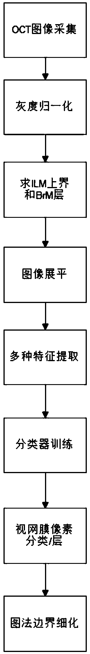
[0038] Such as figure 1 As shown, the specific embodiment of the present invention comprises the following steps:

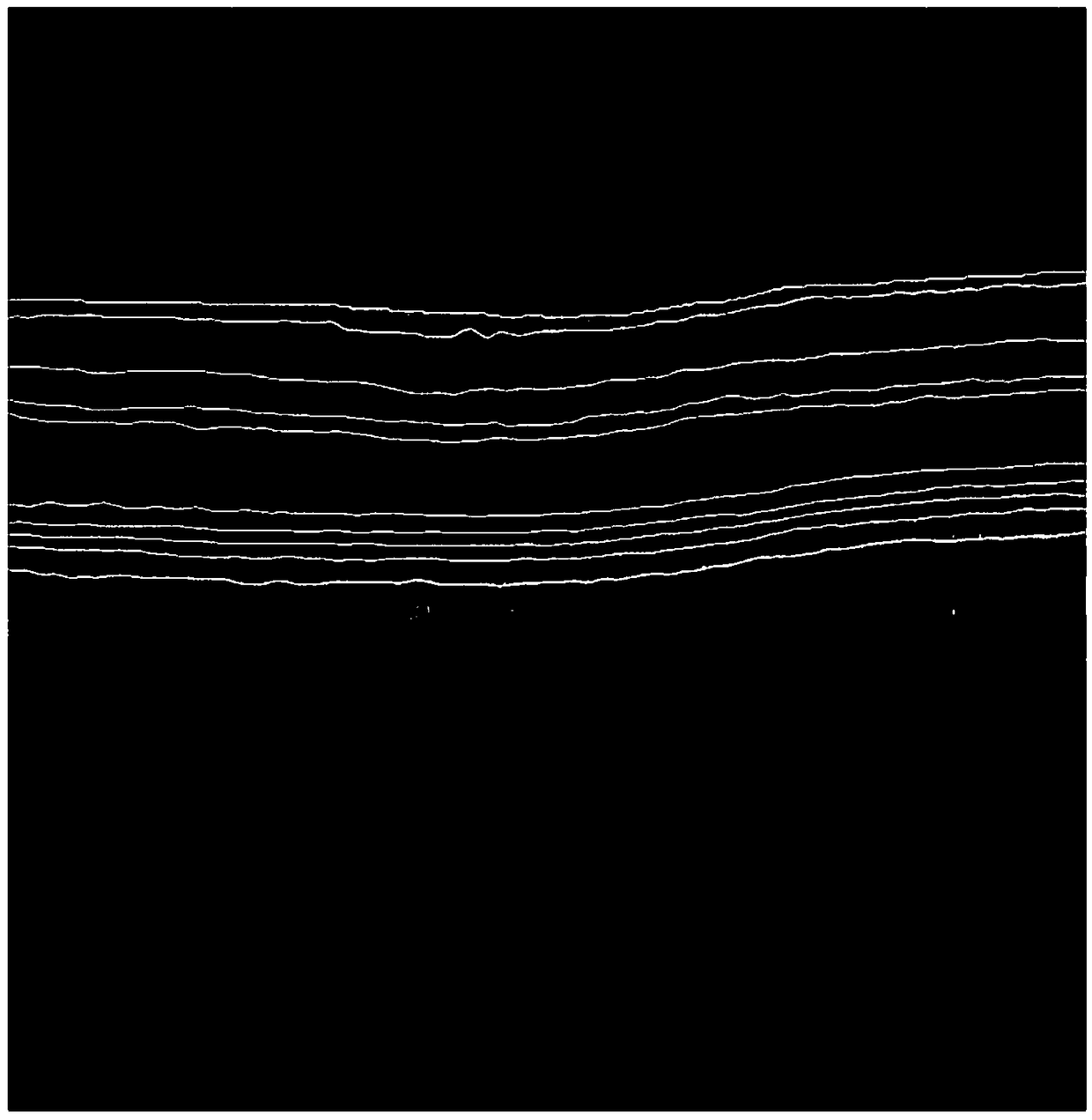
[0039] 1) Collect 40 mouse fundus OCT images as sample images, and the sample image resolution is 1024*1000, such as figure 2 Shown is one of the typical sample images, showing a macular depression with upper ghosting. The image coordinate system is established, with the upper left corner of the image as the origin, the image horizontally to the right as the positive X direction, and the image vertically downward as the positive Y direction.

[0040] 2) Perform grayscale normalization processing on the collected sample images:

[0041] Set the threshold value to 1.05*A, and process the image with the gray value in the range of (0-1.05*A) according to the maximum and minimum normalization processing, and process the pix...


PUM


Abstract

The invention discloses a retinal layering method based on an OCT image. The OCT image of the fundus of the eye is collected as a sample image, the gray-scale normalization processing is carried out on the collected sample images, and then the sample image is flattened. The average filter is used to solve the longitudinal gradient, and then the upper and lower retinal boundaries are determined bycomparison. The calculated features are inputted to the training classifier to obtain the probability that pixels are located at the boundary of different layers. Finally, the optimal boundary and itsposition of the training classifier are optimized by using a graph theory algorithm through the probability. The method of the invention can realize the automatic segmentation of a plurality of layerstructures by using a commonly configured computer, and adopts the multi-level probability classification decision and optimization, and at the same time adopts serialization layering, has fewer original processing steps for the image, and improves detection accuracy certainly.

Description

technical field [0001] The invention belongs to the field of medical image processing, and in particular relates to a retinal layering method based on OCT images. Background technique [0002] OCT interference imaging is based on the optical scattering characteristics of biological tissues to near-infrared light to capture two-dimensional or three-dimensional images, which can achieve micron (μm) resolution. OCT has the advantages of high resolution, strong ease of use, less ionizing radiation, high comfort to patients, and low cost, making it widely used in retinal imaging around the macular cube, and can be used as a basis for auxiliary diagnosis of various diseases. Such as multiple sclerosis (MS), type 1 diabetes, Alzheimer's disease, Parkinson's disease and glaucoma. The above diseases have been shown to have corresponding quantitative abnormalities found in different retinal layers. A better understanding of the impact of these diseases on specific cell types in the ...

Claims


Application Information

Patent Timeline
IPC IPC(8): G06T7/12G06K9/62
CPCG06T7/12G06T2207/30041G06F18/2411G06F18/214
Inventor 周扬岑岗石龙杰周武杰陈正伟陈才毛建卫刘铁兵施秧
Owner ZHEJIANG UNIVERSITY OF SCIENCE AND TECHNOLOGY
Why Eureka
Try Eureka