
3D printed artificial endometrium and its preparation and application

An endometrial, artificial technology, applied in educational appliances, medical science, instruments, etc., can solve the problem that two-dimensional culture conditions cannot fully reproduce the shape, structure and function of the endometrium.

Inactive Publication Date: 2019-02-26
SHENZHEN INST OF ADVANCED TECH
View PDF7 Cites 2 Cited by
  • Summary
  • Abstract
  • Description
  • Claims
  • Application Information

AI Technical Summary

Problems solved by technology

[0013] Regarding the impact of endometrium on embryo implantation, at first, different types of endometrium are usually cultured in vitro to observe their morphological characteristics and responses to different treatment conditions. Although major breakthroughs have been made in related research, but Two-dimensional culture conditions cannot completely reproduce the morphology, structure and function of endometrium in vivo, and there are certain limitations

Method used


Image

  • 3D printed artificial endometrium and its preparation and application
  • 3D printed artificial endometrium and its preparation and application
  • 3D printed artificial endometrium and its preparation and application

Examples


Embodiment 1

[0068] Example 1: Configuration of mixed culture solution

[0069] The composition of the mixed culture medium is: DMEM F12+10% FBS+1% streptomycin+1% penicillin+100nmol / L E2+10nmol / L P4). The mixed culture medium was sterilized by filtration of 0.22 μm at 37°C, 5% CO. 2 Equilibrate in the incubator for 2-4 hours.

Embodiment 2

[0070] Example 2: Acquisition of Endometrial Epithelial and Stromal Cells

[0071] The mouse endometrial tissue was removed under sterile conditions, washed three times with PBS (containing 1% penicillin and streptomycin) to remove blood clots and mucus on the surface of the tissue, placed in a petri dish, and the endometrium was cut into pieces and added 2 ml of 0.3% type I collagenase, mixed well, placed in a water bath at 37°C for 60 min of shaking and digestion; repeated pipetting and mixing, filtered through a 100-mesh filter to remove undigested tissue, and the filtrate was collected; epithelial cells and stromal cells were obtained Crude mixture.

[0072] Separating stromal cells and epithelial cells in the endometrium by centrifugation: centrifuge at 600 r / min for 10 min, and precipitate into epithelial cells; in suspension, stromal cells;

[0073] Centrifuge the stromal cell-rich suspension at 1200r / min for 10min, discard the supernatant, suspend the precipitate with...

Embodiment 3

[0075] Example 3: Preparation of 3D printed endometrium

[0076] Design and preparation of 3D printed endometrial bioscaffolds

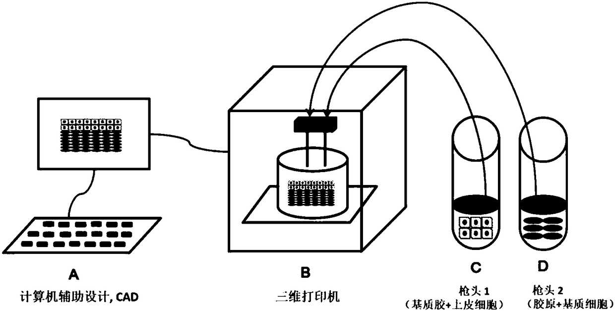
[0077] (1) Combined with Computer Aided Design (CAD), three-dimensional printing technology is used to construct an endometrial stent, in which the shape, composition and internal structure have good designability;

[0078] (2) The shape of the three-dimensional endometrial stent is designed to be similar to the shape of the endometrium, and to ensure that the internal structure is connected and has a certain pore structure;

[0079] (3) The materials used for the three-dimensional endometrial stents were selected to be printed in combination with gelatin, collagen I and matrigel.

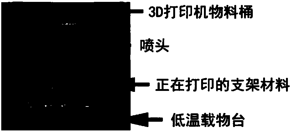
[0080] (4) Internal structure parameters of the constructed three-dimensional endometrium: pore size (R): = 0.2 mm; line stacking angle: 90° (see figure 2 ).

[0081] (5) Store the prepared scaffold at -80°C for later use.

[0082] Preparation of the endometrium

...


PUM

PropertyMeasurementUnit
Diameteraaaaaaaaaa
Pore sizeaaaaaaaaaa

Abstract

The invention relates to an artificial endometrium and its preparation and application and particularly relates to an artificial endometrium. The artificial endometrium comprises a biocompatible stent, endometrial stromal cells / epithelial cells and a mixed culture solution. Through a 3D bioprinting technology, the biocompatible stent and the endometrial stromal cells / epithelial cells are processedinto an artificial endometrium with bioactivity. The mixed culture solution comprises a medium and a biohormone. The biocompatible stent is prepared through a 3D printing method. The artificial endometrium can be used as a research tool for pathophysiological mechanisms aiming at artificial endometrial diseases, can be used as a screening model for regulating and affecting factors related to embryo implantation, and can be used as a research tool for preventing or treating directly or indirectly related endometrial lesions caused by any factor.

Description

technical field [0001] The invention relates to the field of biotechnology, in particular to an artificial endometrium prepared by 3D printing technology. Background technique [0002] The uterus is a part of the female reproductive organs of humans and animals, and it is the place where human and animal fetuses or larvae develop and grow. physiological function. [0003] The endometrium is the mucosal layer in the mammalian uterus, consisting of epithelial cells, stromal cells and blood vessels. The endometrium can be divided into superficial functional membrane and deep basal layer according to its periodic functional changes. / 5. During the menstrual cycle, the functional layer can be peeled off, but the basal layer will not be peeled off. In this process, under the control of ovarian hormones (estrogen and progesterone), the shape and function of the endometrium follow the sexual cycle ( estrus cycle, menstrual cycle) showed obvious morphological and functional change...

Claims


Application Information

Patent Timeline
IPC IPC(8): A61L27/40A61L27/38A61L27/24A61L27/22A61L27/56G09B23/28
CPCA61L27/222A61L27/227A61L27/24A61L27/3813A61L27/3834A61L27/3882A61L27/56A61L2300/412A61L2430/22B33Y10/00B33Y70/00G09B23/28C08L89/00A61L27/22A61L27/38A61L27/40
Inventor 张键阮长顺赵华山任培根杨雅莉孙立峰翟欣昀吴明明
Owner SHENZHEN INST OF ADVANCED TECH
Learn More