
Establishment and evaluation method of animal model for drug in vivo process evaluation after intraperitoneal radiotherapy

A technology of animal models and method establishment, applied in biological testing, instruments, measuring devices, etc., can solve the problem of little research on the interaction between radiotherapy and chemotherapy drugs, and achieve the effect of simple operation and high stability

Active Publication Date: 2022-02-01
CHINA INST FOR RADIATION PROTECTION
View PDF4 Cites 0 Cited by
  • Summary
  • Abstract
  • Description
  • Claims
  • Application Information

AI Technical Summary

Problems solved by technology

[0006] However, at present, normal animals are mostly used to study the in vivo process of drugs, and there are few studies on the interaction between radiotherapy and chemotherapy drugs in combined radiotherapy and chemotherapy. Strengthening the research on the interaction of combined therapy and clarifying the interaction mechanism is the key to formulating a reasonable treatment plan. key

Method used


Image

  • Establishment and evaluation method of animal model for drug in vivo process evaluation after intraperitoneal radiotherapy
  • Establishment and evaluation method of animal model for drug in vivo process evaluation after intraperitoneal radiotherapy
  • Establishment and evaluation method of animal model for drug in vivo process evaluation after intraperitoneal radiotherapy

Examples


Embodiment 1

[0022] Example 1: Establishment of an animal model for drug in vivo process evaluation after abdominal radiotherapy

[0023] The SD rats in the treatment group were weighed and given a single gavage of 500 mg / kg capecitabine, anesthetized with 10% (m / v) chloral hydrate, and fixed on a wooden board. The X-ray simulator was used to locate the irradiated part of the rat between the xiphoid process and the hind limb, and other parts of the rat's body were covered with a lead plate. Rats were irradiated with 6MeV electron beams from a medical linear accelerator at a dose rate of 3Gy / min, and the total single irradiation dose was 10Gy.

Embodiment 2

[0024] Embodiment 2: the evaluation of the animal model established in embodiment 1

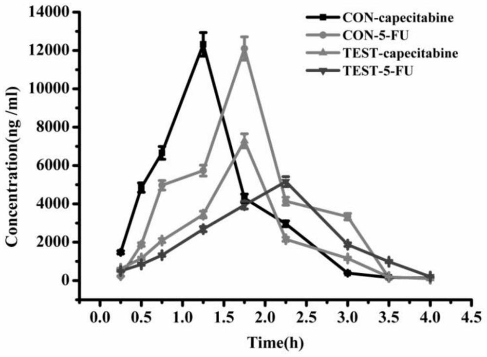
[0025] By means of pharmacokinetics, blood samples were taken from rats at different time points after administration of capecitabine to determine the concentration of capecitabine and its metabolite 5-fluorouracil, and the pharmacokinetic parameters were thus obtained, so as to Established animal models were evaluated. Specific steps are as follows:

[0026] (1) Blood was collected from rats at 0.25, 0.5, 0.75, 1.25, 1.75, 2.25, 3.0, 3.5 and 4.0 hours after administration. Take 400 μL of plasma from each blood sample, add 800 μL of methanol, vortex for 3 minutes, mix well, centrifuge at 4°C and 13000 rpm for 20 minutes, take 10 μL of supernatant, add 90 μL of ultrapure water, and filter with 0.22 μm membrane to prepare test samples for UPLC - MS / MS analytical determination.

[0027] (2) Establish methodology according to the biological sample analysis method, the blood drug concentration ...


PUM


Abstract

The invention belongs to the technical field of animal model construction, and relates to a method for establishing and evaluating an animal model for evaluating the in vivo process of a drug after intraperitoneal radiotherapy. The established method is to irradiate the part between the xiphoid process and the hind limb of the rat with the 5-6MeV electron beam generated by the medical linear accelerator immediately after the chemotherapeutic drug is administered to the rat. The establishment and evaluation method of the animal model for evaluating the drug in vivo process after abdominal cavity radiotherapy of the present invention can make the established rat animal model well simulate the influence of the pathological conditions of the abdominal and pelvic cavity radiation injury caused by clinical radiotherapy on the in vivo process of the chemotherapeutic drug, thereby Contribute to a more rational study of the process of the drug in the patient's body.

Description

technical field [0001] The invention belongs to the technical field of animal model construction, and relates to the establishment and evaluation method of an animal model for evaluating the in vivo process of drugs after abdominal radiotherapy. Background technique [0002] Surgery, chemotherapy and radiotherapy constitute the three pillars of modern medical cancer treatment, and the combination of radiotherapy and chemotherapy has gradually become the first choice for most cancer treatments because of its remarkable curative effect and acceptable side effects. A large number of clinical practices have proved that the calculation of radiotherapy dose is more accurate, making appropriate radiotherapy and chemotherapy plans and more reasonable chemotherapy administration routes can minimize the side effects of radiotherapy and chemotherapy and improve the curative effect. [0003] At present, radiotherapy + capecitabine combination therapy has gradually become the main method...

Claims


Application Information

Patent Timeline
Patent Type & Authority Patents(China)
IPC IPC(8): G01N33/50
CPCG01N33/5008
Inventor 高洁刘欢尹晶晶李建国秦秀军黄立群孙鸽刘梦雅李梅王新钢胡波宋建波杨彪
Owner CHINA INST FOR RADIATION PROTECTION
Learn More