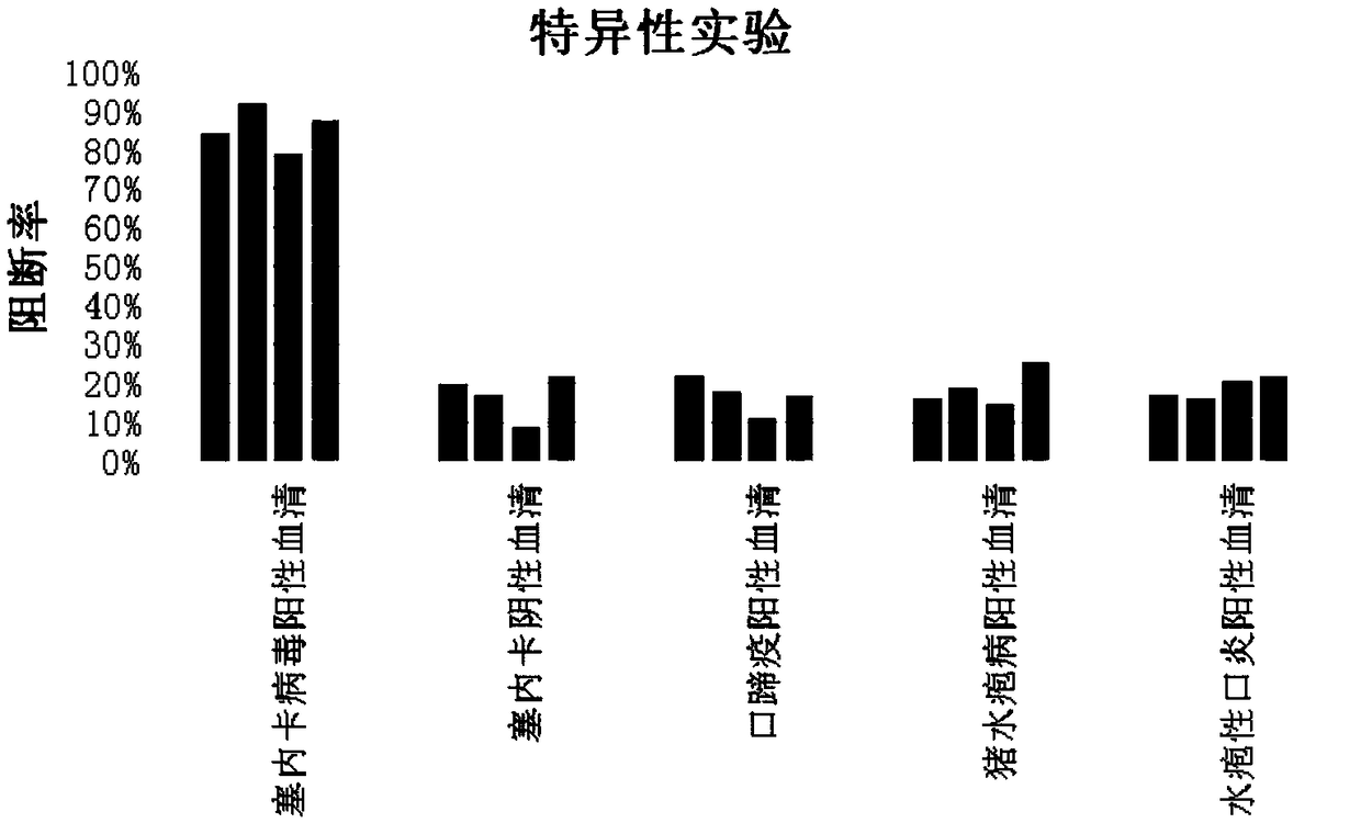
Seneca virus blocking ELISA (enzyme-linked immuno sorbent assay) antibody detection kit
An antibody detection and kit technology, which is applied in the field of Seneca virus antibody detection kits, can solve the problems of complicated operation process and result calculation, high professional technical level requirements, unsuitable pig herd infectivity detection, etc., and achieves good results. The effect of reaction sensitivity, simple and fast preparation process, and loose technical requirements
- Summary
- Abstract
- Description
- Claims
- Application Information
AI Technical Summary
Problems solved by technology
Method used
Image
Examples
Embodiment 1
[0020] Example 1 Preparation of Seneca virus blocking ELISA antibody detection kit
[0021] 1. Preparation of Seneca virus antigen
[0022] Select PK-15 cells (purchased from China Veterinary Drug Control Institute) that are full of monolayers, and inoculate Seneca virus (isolated and identified by the seed virus room of the National Engineering Laboratory of Veterinary Vaccines of Jinyu Baoling Biopharmaceutical Co., Ltd.) at a concentration of 2%. Cultivation), when the CPE (cytopathic change) reaches 85%-90%, harvest the venom, freeze and thaw twice at -20°C, add 6% PEG, stir for 4 hours, let stand overnight, and use 8000rpm / min the next day, Centrifuge for 20 minutes, discard the supernatant, and resuspend the pellet; equilibrate the purification column with equilibrium solution (0.02M disodium hydrogen phosphate, 0.15M sodium chloride, pH7.4) for 5-10 column volumes, and select the sample pump to carry out Load the sample, then use the balance solution to elute, and coll...
Embodiment 2
[0037] The usage method of embodiment 2 Seneca virus blocking ELISA antibody detection kit
[0038] 1. Take the kit obtained in Example 1 out of the refrigerator at 4°C, and return to room temperature after about 15 minutes. Dilute the 20-fold wash solution to 1-fold use concentration with purified water.
[0039] 2. Take out the microtiter plate. The unpacked microtiter plate should be stored at 4°C and should be used within 7 days.
[0040] 3. Use the sample diluent to dilute the serum, and dilute the serum to be tested 10 times. The standard positive serum and standard negative serum do not need to be diluted.
[0041] 4. Adding samples: Add standard positive serum to the 1-2 wells of the first row of the microtiter plate, add standard negative serum to the 3rd to 4th wells, and use the 5th to 6th wells as blank controls. Add the serum to be tested to the remaining wells, add 1 well for each serum, do not repeat, shake gently to mix.
[0042] 5. Incubation: Seal the mic...
Embodiment 3
[0052] Example 3 Repeatability Experiment of Seneca Virus Blocking ELISA Antibody Detection Kit
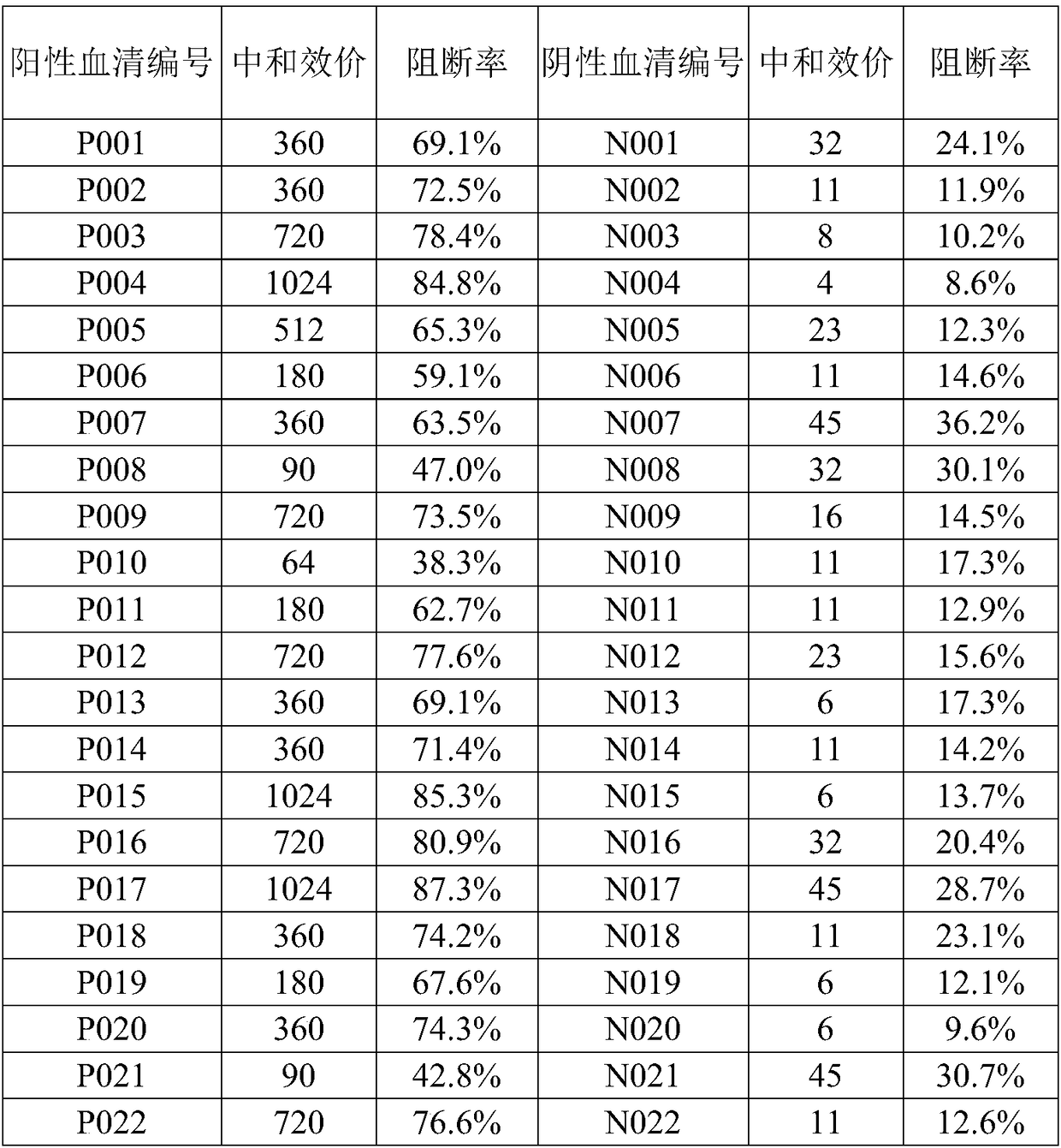
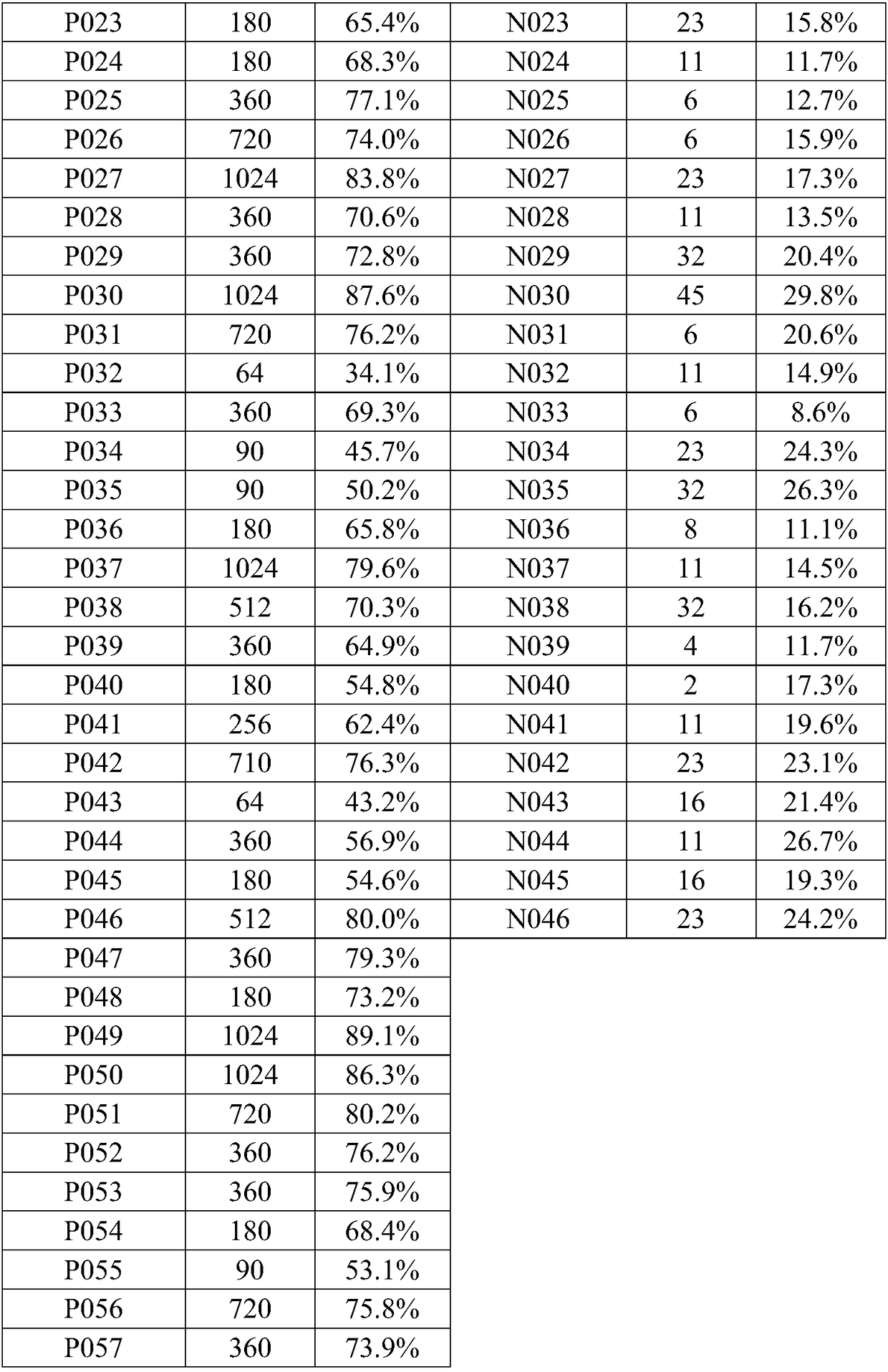
[0053] Select 57 Seneca virus positive sera and 46 negative sera, use the same batch of Seneca virus blocking ELISA antibody detection kit, according to the method of use in Example 2, repeat the detection under the same conditions at different times three times, and the results are shown in Table 1. The results were consistent, indicating that the kits had good reproducibility.
[0054] [Table 1]
[0055]
PUM
 Abstract
Description
Claims
Application Information
 - R&D
- Intellectual Property
- Life Sciences
- Materials
- Tech Scout
- Unparalleled Data Quality
- Higher Quality Content
- 60% Fewer Hallucinations
Browse by: Latest US Patents, China's latest patents, Technical Efficacy Thesaurus, Application Domain, Technology Topic, Popular Technical Reports.
© 2025 PatSnap. All rights reserved.Legal|Privacy policy|Modern Slavery Act Transparency Statement|Sitemap|About US| Contact US: help@patsnap.com


