Method for separating extracellular vesicles based on metal ion complexing agent

A metal ion and separation method technology, applied in the field of biotechnology and nanomaterials, can solve the problems of expensive antibodies and high separation costs, and achieve the effect of easy-to-obtain raw materials, high purity, and simple operation

Inactive Publication Date: 2019-04-05
贺川江
View PDF3 Cites 7 Cited by
  • Summary
  • Abstract
  • Description
  • Claims
  • Application Information

AI Technical Summary

Problems solved by technology

However, due to the high price of antibodies, the separation cost of this method is high

Method used


Image

  • Method for separating extracellular vesicles based on metal ion complexing agent
  • Method for separating extracellular vesicles based on metal ion complexing agent
  • Method for separating extracellular vesicles based on metal ion complexing agent

Examples


Embodiment 1

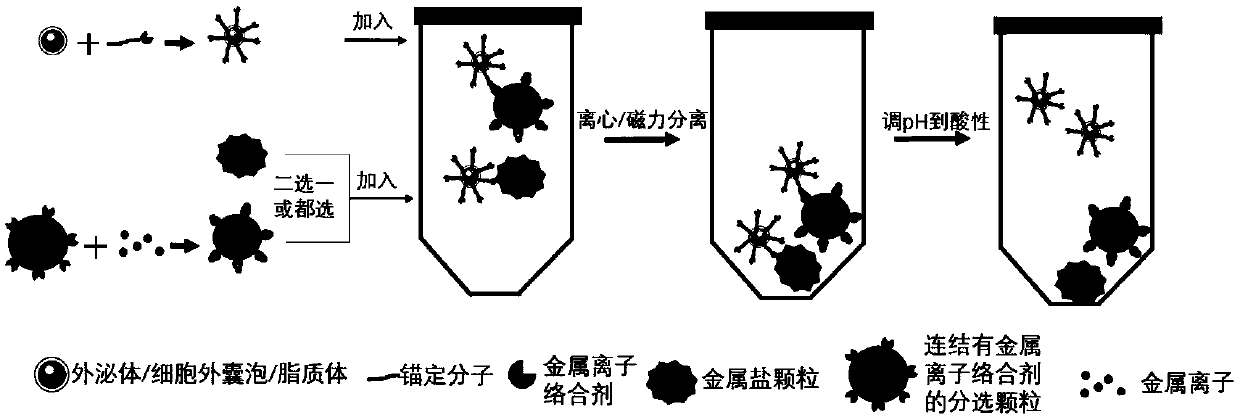
[0045] Separation process such as figure 1 shown. Mix 200 μM oleyl chain molecules modified with amino groups with 200 μM EDTA, react at room temperature for 1 hour, and dilute the reacted mixture to 20 μM. Carry out gradient centrifugation on the cell culture supernatant containing exosomes: centrifuge at 300g for 10min, 2000g for 10min, and 10000g for 30min at 4°C, and take the supernatant after centrifugation. Take the above-mentioned diluted mixed solution and mix it with the supernatant containing the above-mentioned exosomes in equal volumes, and place it at 30°C for 60 minutes of reaction; add the above-mentioned reaction solution into an ultrafiltration tube with a molecular weight cut-off of 100KDa, and centrifuge at a speed of 3000g for 10 minutes. Centrifuge repeatedly 3 times to filter out the oleyl chain molecules and EDTA that are not combined with exosomes. The retained exosomes were resuspended with saline.

[0046] Excess calcium carbonate particles with a ...

Embodiment 2

[0048] Mix 200 μM oleyl chain molecules modified with amino groups with 200 μM EDTA, react at room temperature for 10 hours, and dilute the reacted mixture to 20 μM. Carry out gradient centrifugation on the cell culture supernatant containing exosomes: centrifuge at 300g for 10min, 2000g for 10min, and 10000g for 30min at 4°C, and take the supernatant after centrifugation. Take the above-mentioned diluted mixed solution and mix it with the supernatant containing the above-mentioned exosomes in equal volumes, and place it at 40°C for 10 minutes to react; add the above-mentioned reaction solution to an ultrafiltration tube with a molecular weight cut-off of 80KDa, and centrifuge at a speed of 10,000g for 5 minutes. Centrifuge repeatedly 3 times to filter out the oleyl chain molecules and EDTA that are not combined with exosomes. The retained exosomes were resuspended with saline.

[0049] The magnetic beads coated with iminodiacetic acid (IDA) with a particle size of 100 micron...

Embodiment 3

[0052] Mix 100 μM oleyl chain molecules modified with carboxyl groups with 100 μM glutamic acid diacetic acid (GLDA), react at room temperature for 5 hours, and dilute the reaction mixture to 10 μM. Gradient centrifugation was performed on the urine containing exosomes: centrifuged at 300g for 10min, 2000g for 10min, and 10000g for 30min at 4°C, and the supernatant was taken after centrifugation. Take the above-mentioned diluted mixed solution and mix it with the supernatant containing the above-mentioned exosomes in equal volumes, and place it at 30°C for 60 minutes of reaction; add the above-mentioned reaction solution into an ultrafiltration tube with a molecular weight cut-off of 100KDa, and centrifuge at a speed of 3000g for 10 minutes. Centrifuge repeatedly 3 times to filter out the oleyl chain molecules and GLDA that are not combined with exosomes. The retained exosomes were resuspended with saline.

[0053] Polystyrene microspheres coated with iminodiacetic acid (IDA)...


PUM

PropertyMeasurementUnit
particle diameteraaaaaaaaaa

Abstract

The invention discloses a method for separating extracellular vesicles based on a metal ion complexing agent. The method comprises the steps of performing co-incubation by using an anchoring molecule-metal ion complexing agent molecular complex and liquid which is to be separated and contains the extracellular vesicles, and anchoring the molecular complex onto the extracellular vesicles; mixing sorted particles or salt particles of metal ions with the extracellular vesicles linked with the molecular complex, and incubating to enable the extracellular vesicles to be linked with the sorted particles (or the salt particles); separating, re-suspending a separated product, and adding an eluent to separate the extracellular vesicles from the sorted particles (or the salt particles); and separating out the sorted particles (or the salt particles) to obtain the separated extracellular vesicles remaining in a remaining solution. The method can realize the efficient separation of the extracellular vesicles, does not have special requirements for equipment, and is low in cost and wide in application scope; and exosome obtained by separation is high in purity, and a reagent added in a separating step does not affect the subsequent analysis or application.

Description

technical field [0001] The invention belongs to the field of biotechnology and nanometer materials, in particular to a method for separating extracellular vesicles based on a metal ion complexing agent. Background technique [0002] Extracellular vesicles are a type of lipid vesicles produced by cells that exist outside the cells, including apoptotic bodies, exosomes, and cell budding vesicles. Exosomes are nanovesicles secreted by cells with a lipid bilayer structure, with a particle size distribution of 30-150nm, containing lipids, proteins, nucleic acids and other substances, which are very important in the communication between cells role. Existing studies have confirmed that exosomes are widely involved in various physiological and pathological processes, and can be used as biomarkers for disease screening and diagnosis. Therefore, the isolation and enrichment of exosomes is of great significance in both clinical and scientific research fields. However, because the p...

Claims


Application Information

Patent Timeline
Patent Type & Authority Applications(China)
IPC IPC(8): C12N5/00
CPCC12N5/00C12N2509/00
Inventor 贺川江
Owner 贺川江
Why Eureka
Try Eureka