
medical PET image denoising method based on frequency domain direction smoothing Shearlet

A frequency-domain, smoothing technology, applied in the field of medical PET image denoising, can solve the problems of poor PET image quality, a large amount of hardware noise, software noise and statistical noise electronic devices, and affect the quality of PET images, etc., to achieve good disease analysis and multi-dimensional Singularity approximation, good sparsity effect

Pending Publication Date: 2019-04-05
ZHEJIANG UNIV OF TECH
View PDF5 Cites 1 Cited by
  • Summary
  • Abstract
  • Description
  • Claims
  • Application Information

AI Technical Summary

Problems solved by technology

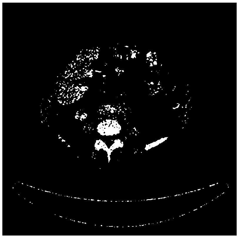
[0004] However, due to the limitations of the PET mechanism imaging, a large amount of hardware noise, software noise, statistical noise, and noise of the electronic device itself will be generated during the imaging process, and most of the PET image noise can be Gaussian white noise with a variance of 0 for its model
The existence of the above noise seriously affects the quality of PET images, resulting in poor PET image quality

Method used


Image

  • medical PET image denoising method based on frequency domain direction smoothing Shearlet
  • medical PET image denoising method based on frequency domain direction smoothing Shearlet
  • medical PET image denoising method based on frequency domain direction smoothing Shearlet

Examples


Embodiment Construction

[0075] The present invention will be further described below in conjunction with accompanying drawing:

[0076] The present invention is based on frequency domain direction smoothing Shearlet medical PET image denoising method, comprises the following steps:

[0077] Step 1) establishes a new medical PET image noise model;

[0078] figure 1 It is that the method of the present invention reads the noisy PET image, and its model is as follows:

[0079] First read the PET file to get the image pixel point r x,y , let the noise-free medical PET image sequence be {r x,y ;x,y=1,2,...,n,n∈N}, where r x,y is the gray value of point (x, y) in the medical PET image. The noise model of noisy medical PET images is generally as follows

[0080] s(x,y)=r(x,y)ε(x,y) (1)

[0081] Here, (x, y) represent the two-dimensional coordinates of the video image, r(x, y) represents the noise-free signal, and ε(x, y) represents the multiplicative noise.

[0082] Logarithmic processing is perform...


PUM


Abstract

The invention discloses a medical PET image denoising method based on frequency domain direction smoothing Shearlet. A new medical PET image Gaussian noise model is provided, then frequency domain multi-scale decomposition and multi-smooth direction decomposition are carried out, new unified threshold processing is carried out on the decomposed high-frequency direction smooth Shearlet coefficient,and then a denoised PET image is generated through inverse Shearlet transformation. Compared with a traditional NSST method (non-subsampled Shearlet transform), the method has the advantages that thedenoising effect is better, the speed is higher, and the method can be better applied to the field of medical PET image denoising.

Description

technical field [0001] The invention is applied to the field of medical PET image denoising, and a method for denoising medical images based on frequency-domain direction smoothing Shearlet transform suitable for medical PET images is designed. Background technique [0002] Positron emission computed tomography (PET) is currently the most advanced medical imaging equipment. Compared with CT and MRI techniques, it has the advantages of high safety, high sensitivity, high specificity, and full-body imaging. It has become one of the most powerful inspection methods in current clinical diagnosis. [0003] Positron emission tomography is a newly developed nuclear medicine examination method. Before scanning, the patient is injected with a radioactive preparation labeled with certain positrons, and the metabolic changes of brain tissue are measured from the metabolic processes they participate in. Since 80% of the energy required by the brain comes from glucose, the more active ...

Claims


Application Information

Patent Timeline
Patent Type & Authority Applications(China)
IPC IPC(8): G06T11/00G06T5/00
CPCG06T11/008G06T2207/10104G06T5/70
Inventor 张聚田峥李澎林周俊赵恺伦
Owner ZHEJIANG UNIV OF TECH
Learn More