Early breast cancer detection medical imaging method based on electromagnetic inverse scattering

A medical imaging and inverse scattering technology, applied in image analysis, image data processing, 3D image processing, etc., can solve problems such as misdiagnosis, long diagnosis and treatment process, etc.

Active Publication Date: 2019-06-07
HANGZHOU DIANZI UNIV
View PDF8 Cites 0 Cited by
  • Summary
  • Abstract
  • Description
  • Claims
  • Application Information

AI Technical Summary

Problems solved by technology

Therefore, this leads to a very long process of diagno

Method used


Image

  • Early breast cancer detection medical imaging method based on electromagnetic inverse scattering
  • Early breast cancer detection medical imaging method based on electromagnetic inverse scattering
  • Early breast cancer detection medical imaging method based on electromagnetic inverse scattering

Examples


Example Embodiment

[0079] Example 1.

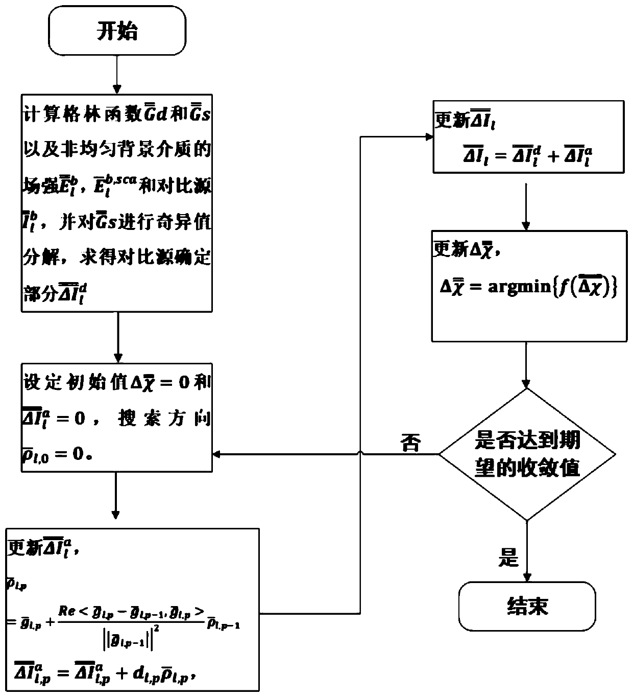
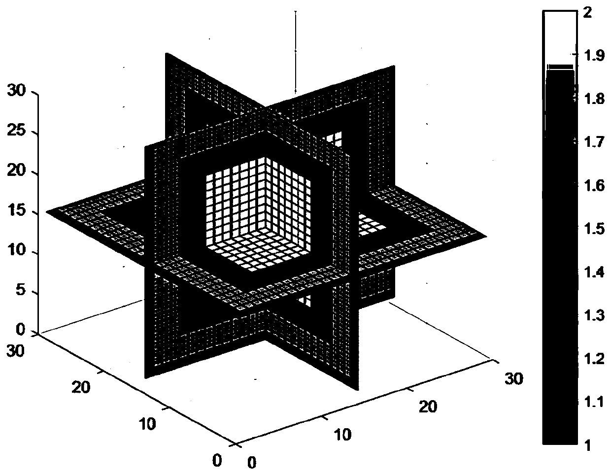
[0080] This example uses experimental data to verify the proposed medical imaging method for early breast cancer detection. The simulation example is a structure composed of a cube culture dish and a group of cube cell groups. The size of the detection area is a cubic area with a side length of 2λ, where λ is the wavelength of the incident wave in space. The background consists of the Petri dish and the free space of the probe area. The side length of the cell body is λ. The length, width and height of the petri dish are both 1.8λ, and the thickness is 0.2λ. It has a relative permittivity of 1.5 and a relative permittivity of 2.0 for cell spheroids. Discretize the entire detection area into a grid of 30×30×30, and use 16 incident points evenly distributed on the 2π solid angle as the emitter, and take the center of the region of interest as the center, in xy, xz, yz three There are 16 receiving devices evenly distributed on the plane, so as to detect th...

Example Embodiment

[0081] Example 2.

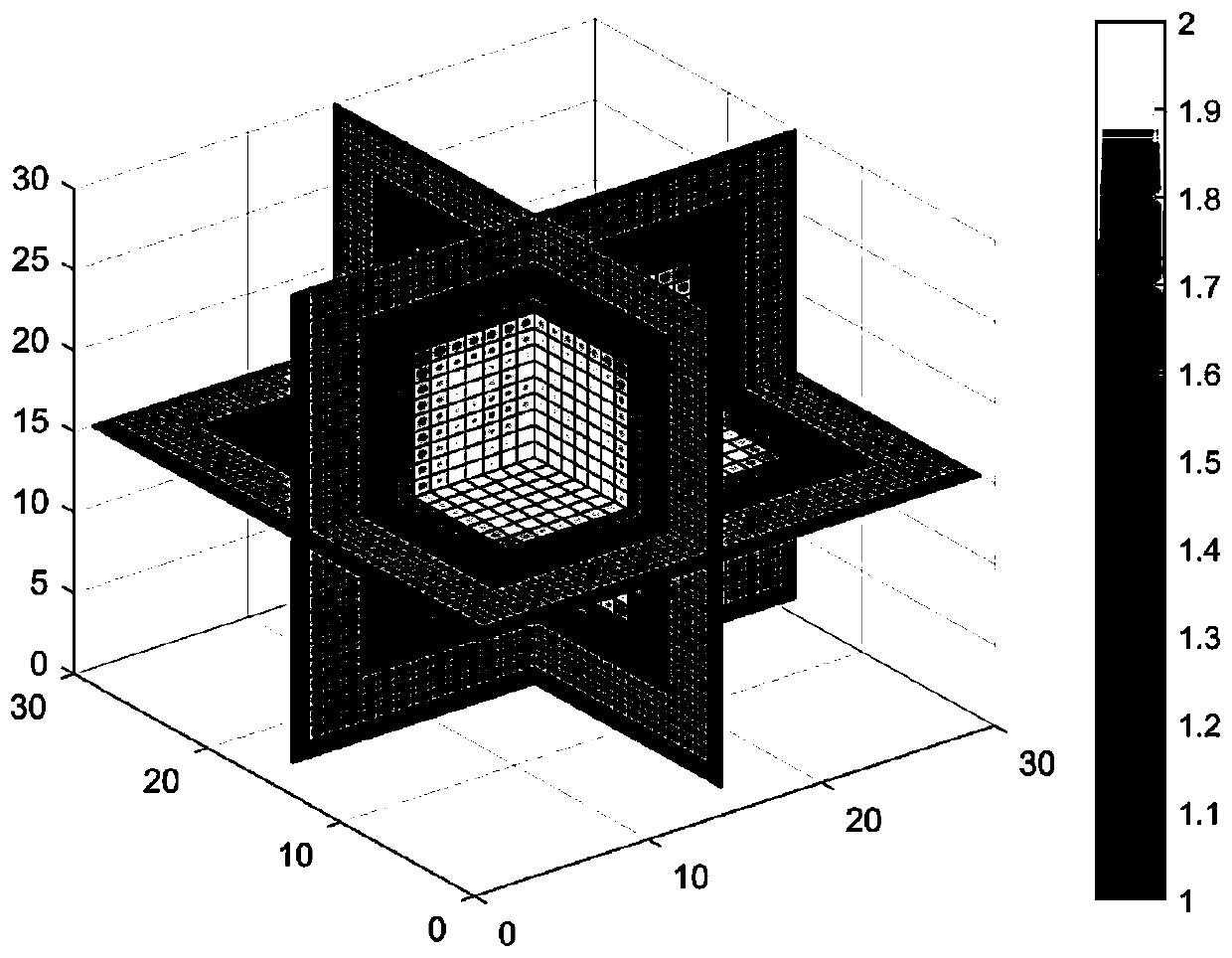
[0082] In order to verify the imaging effect of the early breast cancer detection medical imaging method designed by the present invention on the detection of breast lesion biological tissue, this example 2 carried out inversion imaging to the measurement data of breast tumors, still using the structural device of example 1 to detect breast and Obtain the scattered field data of the detection area, and perform image inversion and reconstruction. Figure 4 is a model device structure for breast tumors, Figure 5 For the reconstruction effect of the breast tumor model, we can clearly find the position, size and dielectric constant of the tumor from the image, which can help the doctor to make an accurate treatment plan. This demonstrates the high accuracy and feasibility of our proposed imaging method.


PUM


Abstract

The invention discloses an early breast cancer detection medical imaging method based on electromagnetic inverse scattering. The method is used for reconstructing information such as the position, theshape and the size of a lesion location which may occur in internal tissues of a living body. After the data information is processed by an algorithm, a visual three-dimensional image can be presented, and the method has the advantages that hospital experts can observe the illness state of the patient more intuitively, and the possibility of artificial misdiagnosis is reduced. According to the scheme, in combination with a subspace optimization method, a novel three-dimensional method based on a differential integral equation model is provided to realize image reconstruction, and in addition,a singular value decomposition and truncation method is used, so that the stability and the accuracy of an iteration process are improved. The result shows that the proposed method not only has verygood convergence performance and fast operation speed, but also can reduce nonlinearity, and can very clearly invert the position size, shape and other information of the biological tissue in the lesion. Test results of multiple experiments verify the effectiveness of the method.

Description

technical field [0001] The invention belongs to the technical field of electromagnetic wave biological imaging, and proposes a medical imaging method for detecting early breast cancer based on electromagnetic inverse scattering. Background technique [0002] In production practice, the three-dimensional imaging method plays a very important role in the process of medical diagnosis and detection of early breast cancer. In the past, we humans often used our own sensory organs, such as eyes, nose, ears, hands, etc., to make pathological diagnoses from the outside of the human body. However, this intuitive feeling, the information obtained is relatively limited. For the exact diagnosis of the disease, especially for the pathological diagnosis inside the body, it is impossible to directly obtain intuitive and accurate information, such as the location, the size of the disease cover, etc., which has great limitations. , only through the human senses is not enough to obtain more u...

Claims


Application Information

Patent Timeline
IPC IPC(8): G06T7/00G06T15/00
Inventor 徐魁文楚彦青叶修竹陈旭东
Owner HANGZHOU DIANZI UNIV
Why Eureka
Try Eureka