Microscopic endoscopy imaging method and system based on structured light illumination

A structured light illumination and imaging system technology, applied in the fields of endoscopy, medical science, diagnosis, etc., can solve the problems of aggravating the financial burden of medical institutions and patients, high light source requirements, complex optical path structure, etc., and achieve fluorescent tissue image information Rich, high fluorescence imaging sensitivity, and high light energy utilization

Pending Publication Date: 2019-08-20
苏州欧谱曼迪科技有限公司
View PDF0 Cites 3 Cited by
  • Summary
  • Abstract
  • Description
  • Claims
  • Application Information

AI Technical Summary

Problems solved by technology

Laser confocal endoscopy has high accuracy and sensitivity in early cancer screening. The disadvantage is that the pinhole device is added, which requires high light source. At the same time, laser confocal technology requires scanning, imaging The speed is slow, and the internal optical path structure is complicated, and the cost is high, which increases the economic burden on medical institutions and patients

Method used


Image

  • Microscopic endoscopy imaging method and system based on structured light illumination
  • Microscopic endoscopy imaging method and system based on structured light illumination
  • Microscopic endoscopy imaging method and system based on structured light illumination

Examples


Embodiment Construction

[0048] The invention discloses a microscopic endoscopic imaging method and system based on structured light illumination, which improves suppression of background light signals and enhances image contrast under the condition of ensuring imaging speed and image resolution.

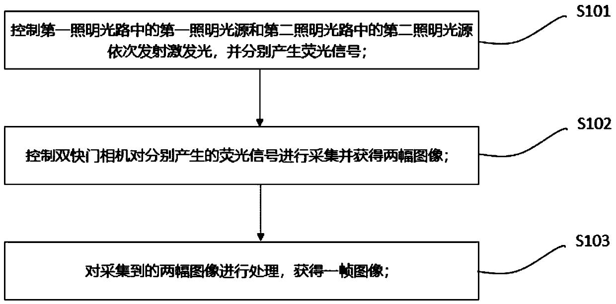
[0049] like figure 1 as shown, figure 1It is a flow chart of the microendoscopic imaging method based on structured light illumination of the present invention. The microendoscopic imaging method based on structured light illumination comprises the following steps:

[0050] S101: Control the first illuminating light source in the first illuminating light path and the second illuminating light source in the second illuminating light path to sequentially emit excitation light, and generate fluorescence signals respectively.

[0051] Specifically, the step of controlling the first illumination light source in the first illumination optical path and the second illumination light source in the second illuminat...


PUM


Abstract

The invention relates to a microscopic endoscopy imaging method and system based on structured light illumination. The method comprises the following steps of controlling a first illumination light source and a second illumination light source to sequentially transmit exciting light, and respectively produce fluorescence signals; controlling a double-shutter camera to acquire the produced fluorescence signals and obtain two images; and performing treatment on the acquired two images to obtain one image, wherein the first illumination light source is a structured light illumination light source, and the second illumination light source is a wide field illumination light source. The system comprises a first illumination light path, a second illumination light path, a beam combining light path and an imaging light path. According to the method and system disclosed by the invention, a scanning device is not needed; a manner of combining structured light illumination image with wide field illumination imaging is adopted, the fluorescence signals are sequentially acquired, the high-quality fluorescence in-focal images can be obtained through synthetizing of the two images, and the imaging speed is high; the situation that a pinhole blocks the light is avoided, the light energy utilization rate is high, the number of the produced fluorescence signals is large, the fluorescence imagingsensitivity is high, and the obtained fluorescence tissue images are rich in information.

Description

technical field [0001] The invention belongs to the field of microscopic endoscopic imaging, in particular to a microscopic endoscopic imaging method and system based on structured light illumination. Background technique [0002] In the field of microendoscopic imaging, optical microscopic imaging has many advantages such as non-destructive, fast, and low-cost, and has been favored by people all the time. Optical microscopic imaging technology is mainly based on the principle of optical microscopy. By using optical lenses to magnify and image objects, the morphological and physiological characteristics of microscopic biological tissue cells can be observed, which is convenient for disease screening and diagnosis of physiological tissues. [0003] High Resolution Fluorescence Microendoscopy (HRME) is a new type of microendoscopy imaging system, which enters the human body through a soft fiber optic bundle, which can simultaneously transmit illumination light and image signal...

Claims


Application Information

Patent Timeline
Patent Type & Authority Applications(China)
IPC IPC(8): A61B1/06A61B1/07A61B1/05A61B1/04
CPCA61B1/043A61B1/05A61B1/0646A61B1/0684A61B1/07
Inventor 杨刚吴郁清林立安昕
Owner 苏州欧谱曼迪科技有限公司
Why Eureka
Try Eureka