Eureka AIR delivers breakthrough ideas for toughest innovation challenges, trusted by R&D personnel around the world.

Two-photon ray-plate microscopic imaging method and device

A technology of microscopic imaging and imaging device, applied in the optical field, can solve the problems of reduced imaging speed, low imaging rate, unable to meet the application requirements of biological dynamic imaging, etc., and achieve the effect of improving imaging efficiency

Active Publication Date: 2019-11-19
TSINGHUA UNIV
View PDF5 Cites 8 Cited by
  • Summary
  • Abstract
  • Description
  • Claims
  • Application Information

AI Technical Summary

Problems solved by technology

However, it is worth noting that the conventional method uses the method of moving the objective lens or sample in the axial direction of the excitation objective lens for scanning, and its mechanical inertia results in a low imaging rate, which cannot meet the application requirements of biological dynamic imaging.
Although people use fast focusing systems, such as electrically tunable lenses or ultrasonic tunable lenses (Cell Research 25, 254–257 (2015)), to increase the scanning speed of the excitation objective lens in the axial direction, the above methods can only perform continuous scanning
However, for some biological dynamic observations in the axial direction of the excitation objective lens, only the region of interest needs to be observed. At this time, continuous scanning will not only reduce the imaging speed, but will also increase the photodamage of the tissue.
[0005] Two-photon light sheet imaging usually uses push-broom linear excitation combined with (virtual) confocal detection to achieve imaging, but the mechanical inertia of the scanning device severely limits the imaging speed. Therefore, it is an urgent problem for those skilled in the art to solve The technical problem is: how to design a two-photon light-sheet microscopic imaging system that scans rapidly in the axial direction of the excitation objective lens and is selective for the target excitation field of view

Method used


Image

  • Two-photon ray-plate microscopic imaging method and device
  • Two-photon ray-plate microscopic imaging method and device
  • Two-photon ray-plate microscopic imaging method and device

Examples


Embodiment Construction

[0053] Embodiments of the present invention are described in detail below, examples of which are shown in the drawings, wherein the same or similar reference numerals designate the same or similar elements or elements having the same or similar functions throughout. The embodiments described below by referring to the figures are exemplary and are intended to explain the present invention and should not be construed as limiting the present invention.

[0054] The two-photon light sheet microscopic imaging method and device proposed according to the embodiments of the present invention will be described below with reference to the accompanying drawings.

[0055] Firstly, a two-photon light sheet microscopic imaging method proposed according to an embodiment of the present invention will be described with reference to the accompanying drawings.

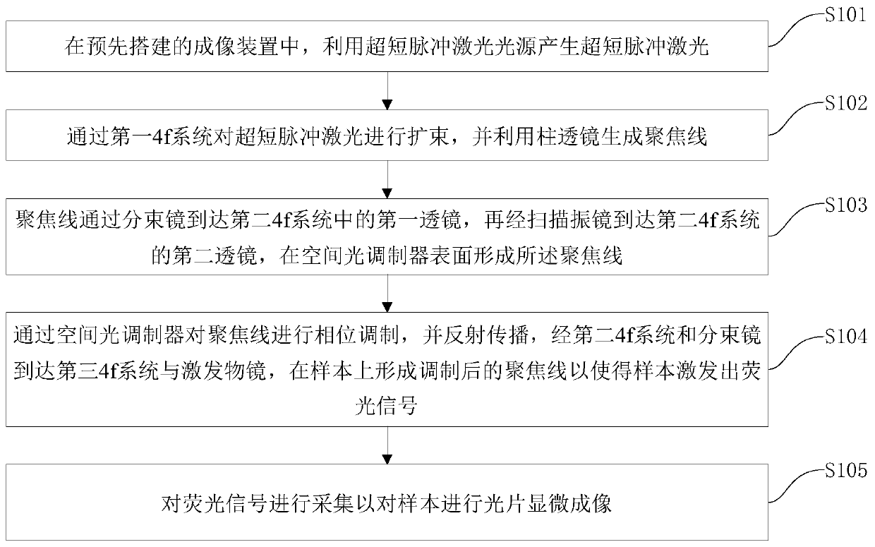
[0056] figure 1 It is a flowchart of a two-photon light sheet microscopic imaging method according to an embodiment of the present inv...


PUM


Abstract

The invention discloses a two-photon ray-plate microscopic imaging method and device. The method comprises the following steps: producing ultra-short pulse laser by utilizing ultra-short pulse laser source in a pre-constructed imaging device; performing beam expanding on the ultra-short pulse laser through a first 4f system, and generating a focusing line by utilizing a cylindrical lens, wherein the focusing line arrives a first lens in a second 4f system through a beam splitter, and then arrives a second lens of a second 4f system through a scanning galvanometer, thereby forming the focusingline on a surface of a spatial light modulator; performing phase modulation on the focusing line through the spatial light modulator, and then reflecting and propagating the focusing line, thereby arriving a third 4f system and an excitation objective lens through the second 4f system and the beam splitter; and collecting a fluorescence signal so as to perform ray-plate microscopic imaging of a sample. Through the method disclosed by the invention, the quick imaging is realized by quickly accessing modulated phase on the spatial light modulator, and the imaging speed and the imaging efficiencyare improved.

Description

technical field [0001] The invention relates to the field of optical technology, in particular to a two-photon light sheet microscopic imaging method and device. Background technique [0002] Light Sheet Microscopy (LSM) is a new type of optical microscopy imaging method. Compared with confocal fluorescence microscopy, light sheet microscopy greatly reduces photobleaching and improves imaging data throughput. In recent years, It has been widely used in biomedical microscopy imaging. [0003] However, considering the influence of biological tissue scattering and other factors, conventional light sheet microscopy based on single-photon excitation has shallow penetration depth and low imaging signal-to-noise ratio, which limits the wide application of this technology. Two-photon fluorescence excitation is based on the nonlinear effect of two-photon absorption. Its excitation wavelength is twice the wavelength of the corresponding single-photon excitation light, thereby reduci...

Claims


Application Information

Patent Timeline
IPC IPC(8): G01N21/64G01N21/01
CPCG01N21/01G01N21/6428G01N21/6456G01N21/6458G01N2021/0112
Inventor 孔令杰张亿季向阳戴琼海
Owner TSINGHUA UNIV
Why Eureka
Eureka Blog
Learn More