P53 protein and mitochondria double-labeled immunofluorescence detection method and kit thereof

A technology for immunofluorescence detection and mitochondria, which is applied in the fields of biomedicine and biology, can solve the problems of different organelles and different target protein localization processes, and achieve the effect of strong sensitivity, good specificity and good stability

Active Publication Date: 2019-12-20
WEST CHINA HOSPITAL SICHUAN UNIV
View PDF5 Cites 3 Cited by
  • Summary
  • Abstract
  • Description
  • Claims
  • Application Information

AI Technical Summary

Problems solved by technology

[0003] At present, there are still many difficulties in the localization of proteins in cells, manifested in: different organelles, the localization process of the corresponding targeting protein is also different, some proteins are not fixed in a single subcellular location, but dynamically in multiple transport between subcellular locations

Method used


Image

  • P53 protein and mitochondria double-labeled immunofluorescence detection method and kit thereof
  • P53 protein and mitochondria double-labeled immunofluorescence detection method and kit thereof
  • P53 protein and mitochondria double-labeled immunofluorescence detection method and kit thereof

Examples


Embodiment 1

[0101] (1) Prepare cell slides and wash:

[0102] A) Preparation of cell slides:

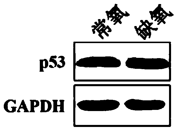
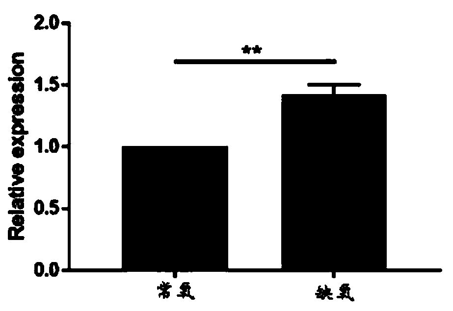
[0103] Cells in the normoxia group: Place 14mm cell slides (NEST) in a 24-well plate, coat with PDL for 5 minutes, wash twice with sterile deionized water and dry naturally; digest the cells and resuspend them, press 5x 10 4 Add it to the well plate according to the calculation per well, shake it crosswise and put it in the incubator for static culture. After 24 hours of culture, confirm that the cells are adhered to the wall and fully stretched out.

[0104] Hypoxia group cells: 14mm cell slides (NEST) were placed in a 24-well plate, coated with PDL for 5 minutes, washed twice with sterile deionized water and dried naturally; the cells were digested and resuspended, and weighed 5x 10 4 Calculated per well and added to the well plate, after cross-shaking, put it into a 1% oxygen concentration tri-gas incubator and culture it in hypoxia for 24 hours.

[0105] B) cleaning:

[0106] Rinse slides...

Embodiment 2

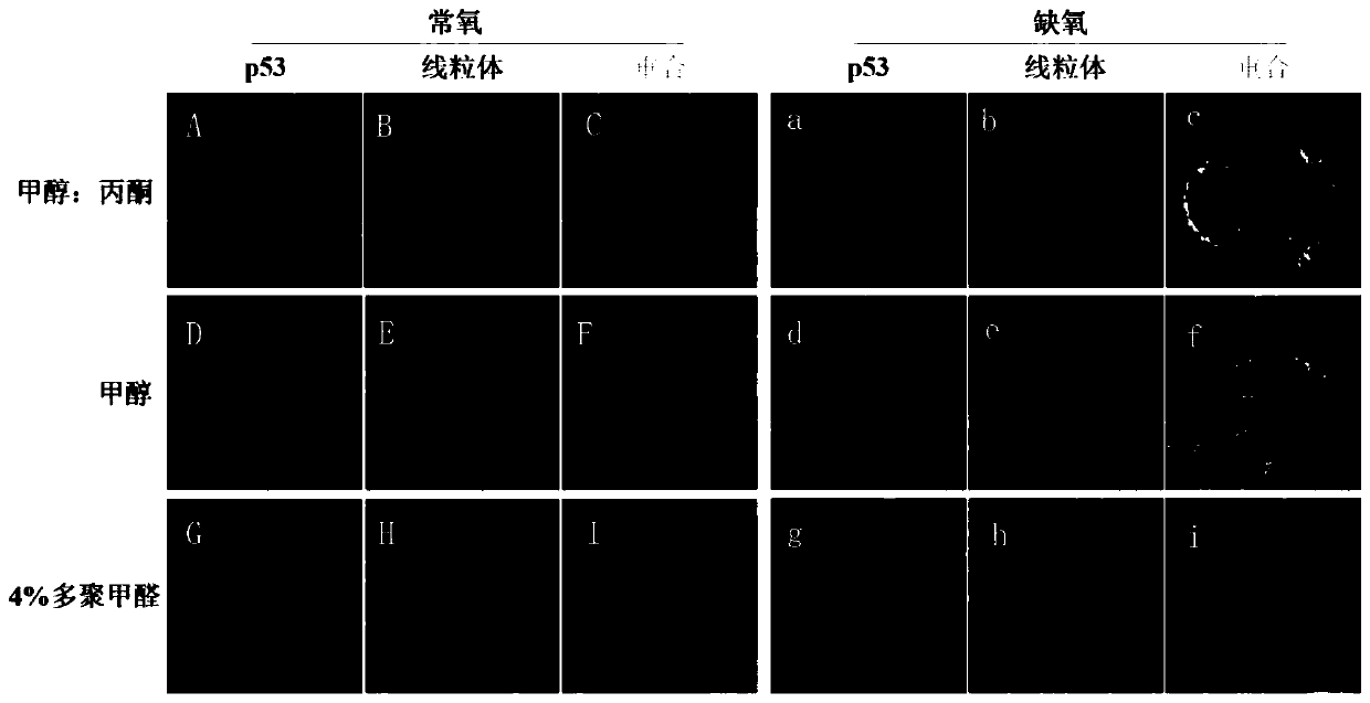
[0115] The only difference with Example 1 is:

[0116] Step (2): add methanol to fix at 4°C for 10 minutes, wash with PBS 3 times, 10 minutes each time.

Embodiment 3

[0118] The only difference with Example 1 is:

[0119] Step (2):

[0120] A) Add 4% paraformaldehyde and fix at 10-30°C for 10 minutes, wash with PBS 3 times, 10 minutes each time;

[0121] B) Permeabilize with 0.1% Triton-X 100, wash 3 times with PBS, 10 minutes / time;

[0122] Step (3): Place the cell slide on a glass slide, put it in a wet box, and block it with 1% BSA at 37° C. for 30 minutes.


PUM


Abstract

The invention discloses a p53 protein and mitochondria double-labeled immunofluorescence detection method and a kit thereof. The detection method comprises the steps of: preparing a cell climbing piece, and performing cleaning; fixing the cell climbing piece with cell fixing liquid, and performing cleaning; sealing the cell climbing piece with cell confining liquid; adding primary antibodies for incubation, and performing rinsing, wherein the primary antibodies comprise an antibody combined with p53 protein and an antibody combined with mitochondria; adding a fluorescently labeled secondary antibody for dyeing, and performing incubation and cleaning; performing redyeing by using cell nucleus fluorochrome, and performing cleaning; sealing the piece, and observing a dyeing result via microscopic examination of a fluorescence microscope or via the microscopic examination of a confocal microscopy; and performing superposed analysis on fluorescent pictures. Only a cytomembrane is broken, anuclear membrane is not broken, intranuclear signal interference can be avoided, co-localization of p53 protein and mitochondria is realized, and basis and reference are provided for the positioning of the mitochondria of protein and the positioning study of the protein and other non-nuclear organelles.

Description

technical field [0001] The invention relates to the fields of biomedicine and biotechnology, in particular to a p53 protein and mitochondria double-labeled immunofluorescence detection method and a kit thereof. Background technique [0002] Eukaryotic cells have a complex subcellular structure, and dozens of organelles have been found, each of which has a specific set of proteins. In eukaryotic cells, except chloroplasts and mitochondria, which can synthesize a small amount of proteins, most of the proteins are synthesized in the cytoplasm or rough endoplasmic reticulum, and finally transported to different locations to form mature proteins and perform functions. The localization of proteins in cells is a central issue in cell biology research and a hot topic in molecular biology research. [0003] At present, there are still many difficulties in the localization of proteins in cells, manifested in: different organelles, the localization process of the corresponding targeti...

Claims


Application Information

Patent Timeline
Patent Type & Authority Applications(China)
IPC IPC(8): G01N33/533G01N33/68
CPCG01N33/533G01N33/6803
Inventor 龙丹周彦妮冯莉陈雪璐李胜富
Owner WEST CHINA HOSPITAL SICHUAN UNIV
Why Eureka
Try Eureka