
Intracranial aneurysm detection method and system based on convolutional neural network

A convolutional neural network and intracranial aneurysm technology, applied in the field of medical image processing, can solve the problems of less medical data, low sensitivity, and heavy workload of intracranial aneurysms

Active Publication Date: 2020-02-21
HUAZHONG UNIV OF SCI & TECH
View PDF5 Cites 9 Cited by
  • Summary
  • Abstract
  • Description
  • Claims
  • Application Information

AI Technical Summary

Problems solved by technology

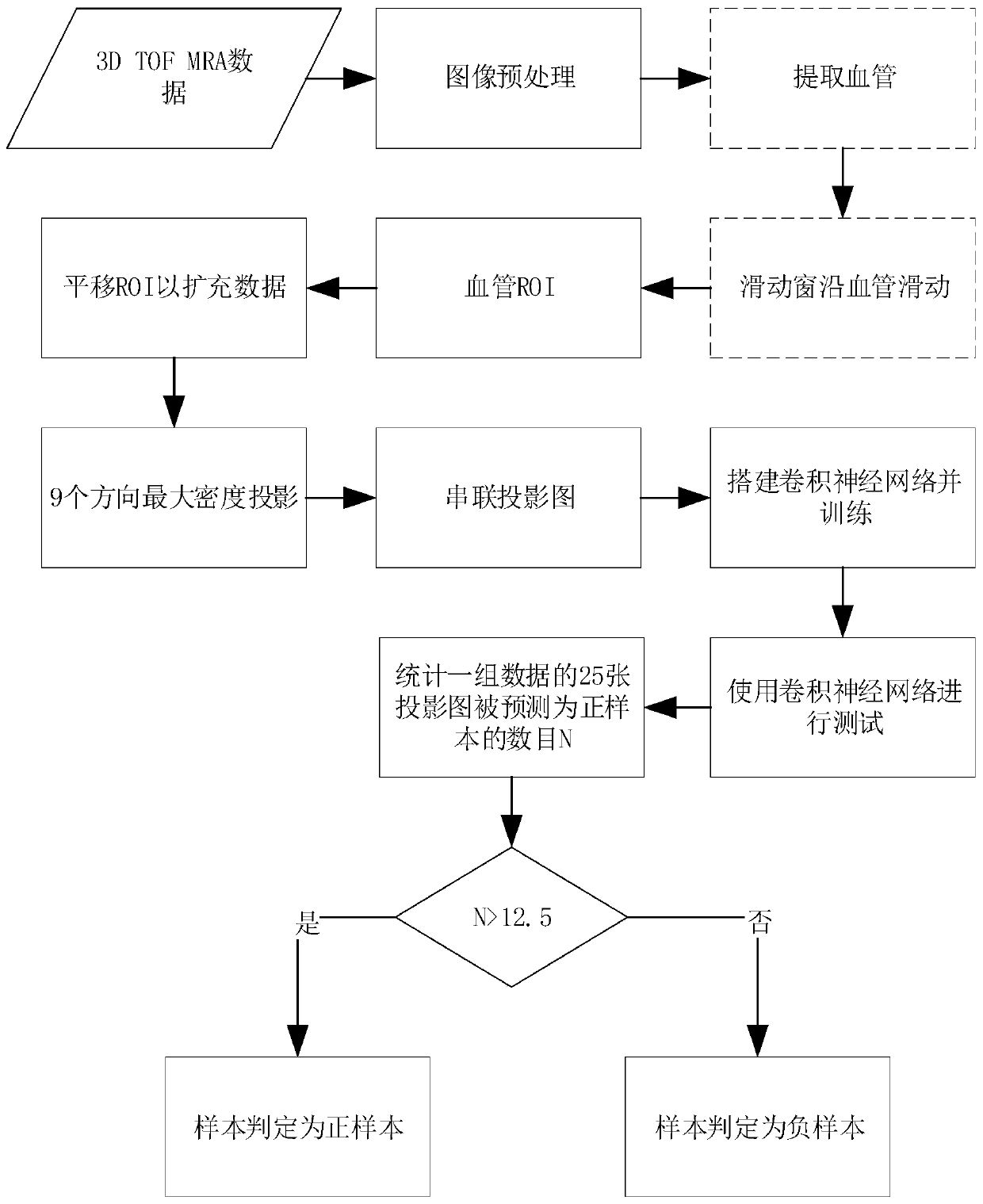
[0004] In view of the problems in the prior art that doctors diagnose intracranial aneurysms from images have a large workload and low sensitivity, the purpose of the present invention is to provide a method and system for detecting intracranial aneurysms based on convolutional neural networks. Improve the overall flow process of the method and the setting method of each functional module component in the corresponding system device. Based on the three-dimensional time-flight magnetic resonance angiography data, first extract the blood vessel, and then use the centerline of the blood vessel as the sliding route to slide and select the cube The voxel block is used as the region of interest ROI, and the maximum density projection is performed on the cube voxel block in multiple directions to obtain the MIP map; by introducing the MIP map into the identification of aneurysms, that is, using the MIP map as input and using the trained The convolutional neural network classifies the MIP map, and the obtained classification result can reflect whether there is an aneurysm in the ROI cube voxel block, and then judge whether there is an intracranial aneurysm in the object to be detected. The detection method and system It has high classification accuracy and sensitivity
In addition, the present invention effectively solves the problem of less medical data by expanding the training positive and negative samples used in the training set

Method used


Image

  • Intracranial aneurysm detection method and system based on convolutional neural network
  • Intracranial aneurysm detection method and system based on convolutional neural network
  • Intracranial aneurysm detection method and system based on convolutional neural network

Examples


Embodiment 1

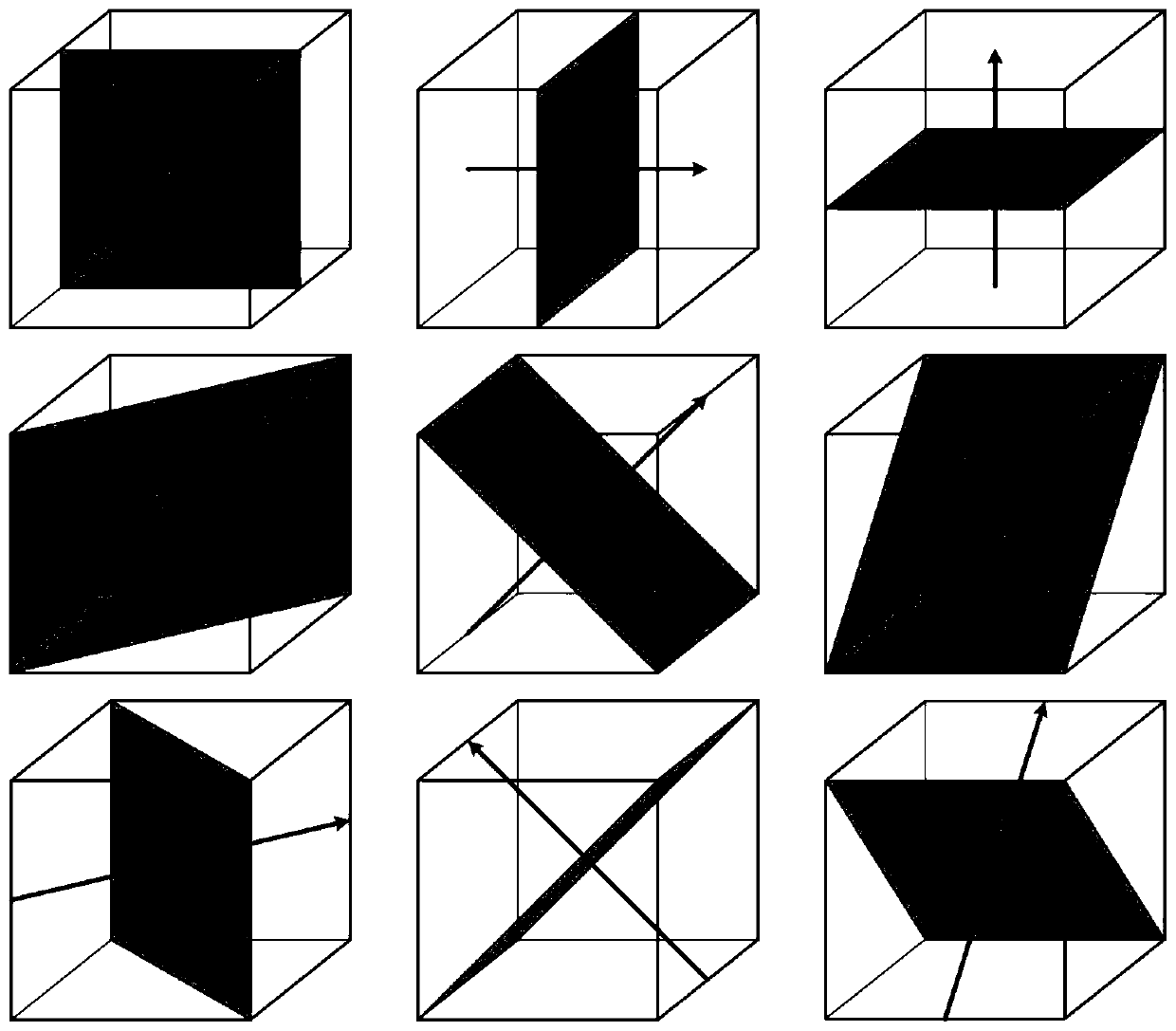
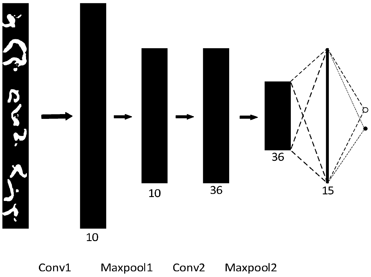
[0072] Generally speaking, when the present invention classifies aneurysms, it first extracts the blood vessels to obtain the position information of the blood vessels, and then uses the position information of the blood vessels to slide and select small cube blocks with the centerline of the blood vessels as the sliding route. Blocks are subjected to maximum density projection in nine directions to obtain a MIP map. Using the MIP map as input, the two-dimensional convolutional neural network is used to classify the MIP map, and the classification result is obtained. The classification result reflects whether there is an aneurysm in the cube block.

[0073] Such as figure 1 As shown, the intracranial aneurysm recognition method based on the MIP image and the convolutional neural network in the embodiment of the present invention includes the following steps:

[0074] (1) Perform preprocessing such as grayscale stretching and normalization on the three-dimensional time-flight ...


PUM


Abstract

The invention belongs to the field of medical image processing. The invention discloses an intracranial aneurysm detection method and system based on a convolutional neural network. The detection method is based on a three-dimensional time leap magnetic resonance angiography image (3D Time-of-Flight MR Angiography, 3D TOF MRA), and is characterized in that first of all, blood vessels are extracted, a series of cubic voxel blocks are extracted as ROIs (Region of Interest) along the central line of the blood vessel; maximum density projection is performed on each ROI (Region of Interest) in a plurality of directions to obtain a MIP (Maximal Intensity Projection) by taking the MIP as the ROI; the MIP graph is used as input, the trained convolutional neural network is used for classifying theMIP graph, an obtained classification result reflects whether the ROI contains the aneurysm or not, and then whether the intracranial aneurysm exists in the to-be-detected object or not is judged. According to the method and the system, the whole process of the method is processed, and the setting mode of each functional module component in the corresponding system device is improved, so that thedetection method and the detection system have relatively high classification accuracy and sensitivity.

Description

technical field [0001] The invention belongs to the field of medical image processing, and more specifically relates to a method and system for detecting an intracranial aneurysm based on a convolutional neural network. Background technique [0002] Intracranial aneurysms are typical of cerebrovascular disease, and their rupture usually results in severe neurologic sequelae and can be fatal. Clinically, three-dimensional time-flight magnetic resonance angiography has been widely used as a screening method for cerebral aneurysms. However, visual detection of unruptured intracranial aneurysms in 3D time-flight magnetic resonance angiography images is difficult for radiologists. [0003] When doctors diagnose intracranial aneurysms clinically, they can observe MIP images of blood vessels at various angles through Magnetic Resonance Imaging (MRI) monitors to determine whether intracranial aneurysms really exist in blood vessels. However, physician fatigue leads to the potentia...

Claims


Application Information

Patent Timeline
IPC IPC(8): G06T7/00G06T17/00
CPCG06T7/0012G06T17/00G06T2207/10088G06T2207/20104G06T2207/20081G06T2207/20084G06T2207/30101G06T2207/30096
Inventor 侯文广邹应诚梅少杰邓先波
Owner HUAZHONG UNIV OF SCI & TECH
Learn More