Application of FTY720-Phosphate in preparation of activating pharmacy of TREM2
A technology of myeloid cells and receptors, applied in the field of drug research and development, to achieve the effect of enhancing phagocytosis and promoting clearance
- Summary
- Abstract
- Description
- Claims
- Application Information
AI Technical Summary
Problems solved by technology
Method used
Image
Examples
Embodiment 1
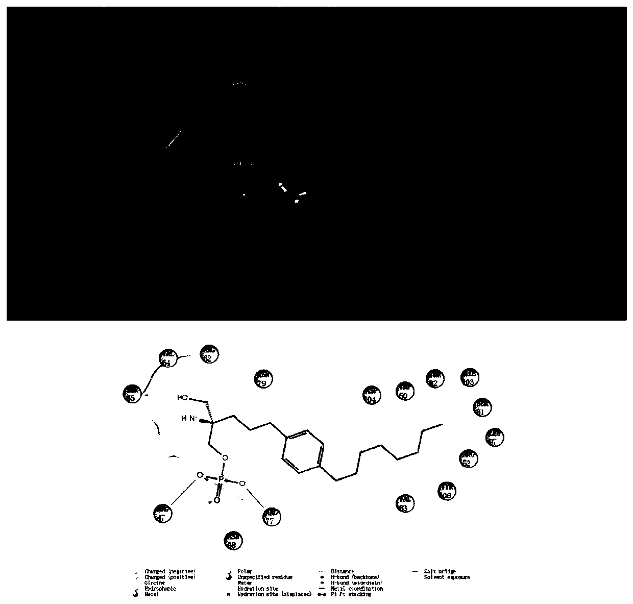
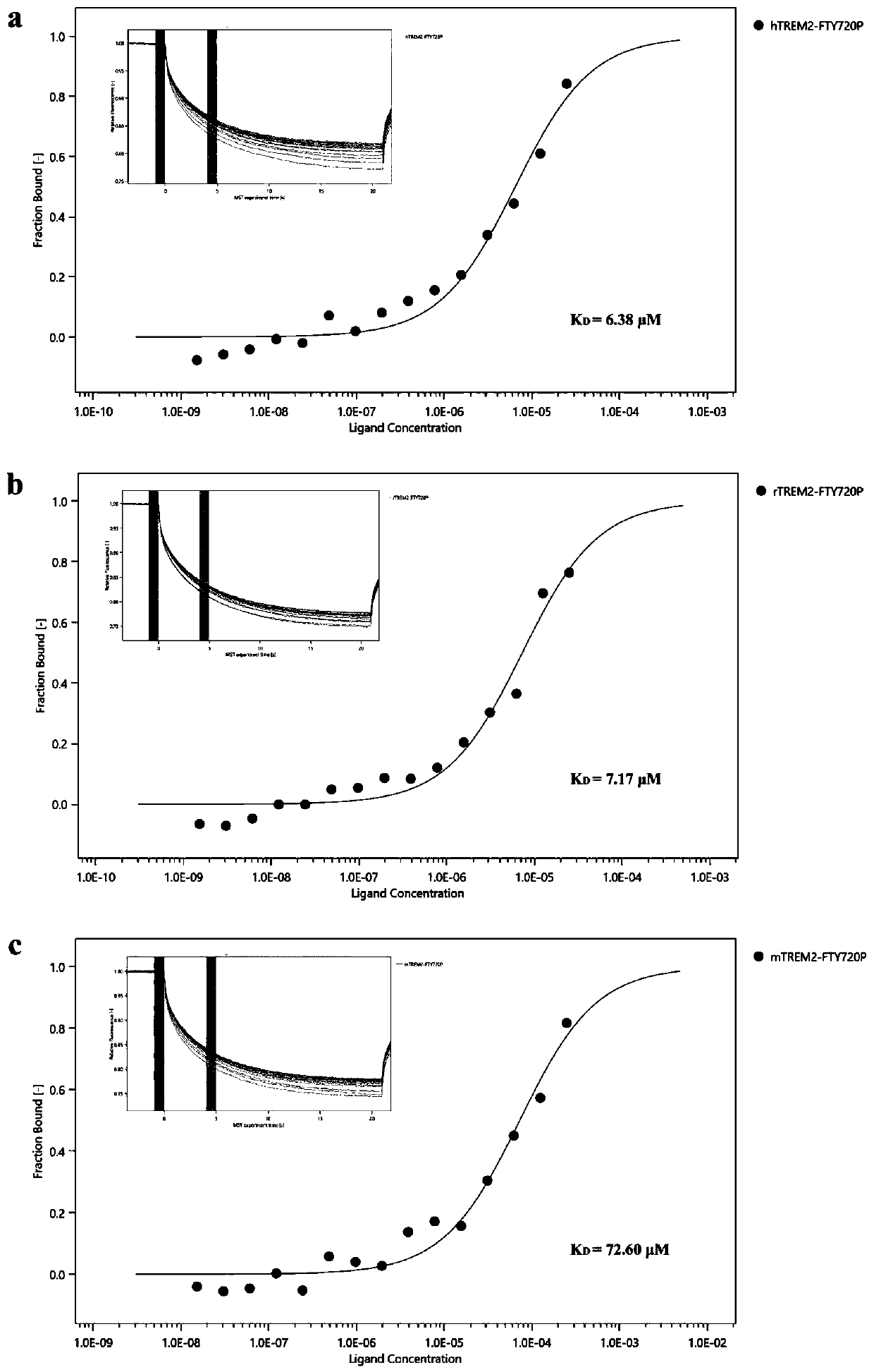
[0026] Microthermal Surge Method Reveals the Relationship Between FTY720-P and TREM2
[0027] Using pET-24a(+) as a vector, construct TREM2 protein expression plasmids of various origins marked with a C-terminal His-tag. The sequences used for human-derived, rat-derived and mouse-derived TREM2 are shown in SEQ ID NO: 1. Shown in SEQ ID NO: 2, SEQ ID NO: 3.
[0028] Transform the recombinant plasmid into BL(DE3) Escherichia coli: Take 100 μL of freshly prepared competent BL(DE3) cells, add 10 μL of the recombinant plasmid, flick and mix well, place on ice for 30 minutes; heat shock at 42°C for 80 seconds, and quickly transfer to ice Cool in the bath for 5 minutes; add 400 μL of LB medium at 37°C, transfer to the incubator, and incubate at 37°C for 1 hour to recover the cells; take an appropriate amount of transformation products and spread them on LB plates containing kanamycin (agar 15g, Peptone 10g, yeast extract 5g, NaCl 10g, water 1000mL), 37 ℃ incubator upside-down cultur...
Embodiment 2
[0031] Experimental grouping and drug treatment
[0032] Rat primary neurons and microglia were extracted respectively, and the primary neurons were cultured in 24-well plates (200,000 cells / well) with Neurobasal medium + 2% B27 + 1% double antibody, and the medium was half changed every other day. Culture for one week until cell maturity; primary microglial cells were cultured with 10% Gibco FBS+DMEM+1% double antibody, and the medium was changed every three days, and culture for one week until cell maturity.
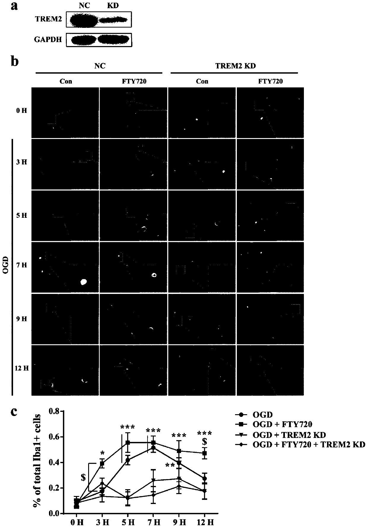
[0033]After the cells matured, microglia were added to neurons for co-culture (20,000 cells / well), co-cultured at a ratio of 1:10, and replaced with Neurobasal medium without B27: (10% Gibco FBS+DMEM+1% double antibody)=3:1 medium culture, divided into normal group, normal drug group, normal knockdown group, knockdown drug group, and each time point after oxygen glucose deprivation-reperfusion (OGD / R) injury These four groups of (3h, 5h, 7h, 9h, 12h). The specific op...
Embodiment 3
[0035] Immunofluorescence staining test
[0036] After the cells were fixed, they were washed 3 times with 0.01M PBS, discarded, and then blocked by adding blocking solution (containing 5% goat serum and 0.1% Triton X-100) at room temperature for 1 hour; adding the primary antibody dropwise (see Table 1 for antibody titer), 4 Incubate overnight at °C. After taking it out the next day, wash the primary antibody with 0.01MPBS, wash 3 times, 5min each time, add the corresponding fluorescent secondary antibody dropwise (see Table 2 for antibody titer), incubate at room temperature for 1h in the dark, and then wash 3 times with 0.01M PBS , 5 min each time; add 5 μg / mL Hoechst solution dropwise to react in the dark for 20 min, wash 3 times with 0.01M PBS, 5 min each time, and take pictures.
[0037] Table 1 Primary antibody for immunofluorescence staining
[0038]
[0039] Table 2 Secondary antibody for immunofluorescence staining
[0040]
[0041] The result is as image ...
PUM
 Abstract
Description
Claims
Application Information
 - R&D
- Intellectual Property
- Life Sciences
- Materials
- Tech Scout
- Unparalleled Data Quality
- Higher Quality Content
- 60% Fewer Hallucinations
Browse by: Latest US Patents, China's latest patents, Technical Efficacy Thesaurus, Application Domain, Technology Topic, Popular Technical Reports.
© 2025 PatSnap. All rights reserved.Legal|Privacy policy|Modern Slavery Act Transparency Statement|Sitemap|About US| Contact US: help@patsnap.com


