

Histopathological tissue sample treatment method
A technology for tissue samples and processing methods, applied in sampling, scientific instruments, preparation of samples for testing, etc., can solve problems such as difficulty in pathological diagnosis, and achieve the effect of maintaining color stability time, good staining effect, and high stability
- Summary
- Abstract
- Description
- Claims
- Application Information
AI Technical Summary
Problems solved by technology
Method used
Image

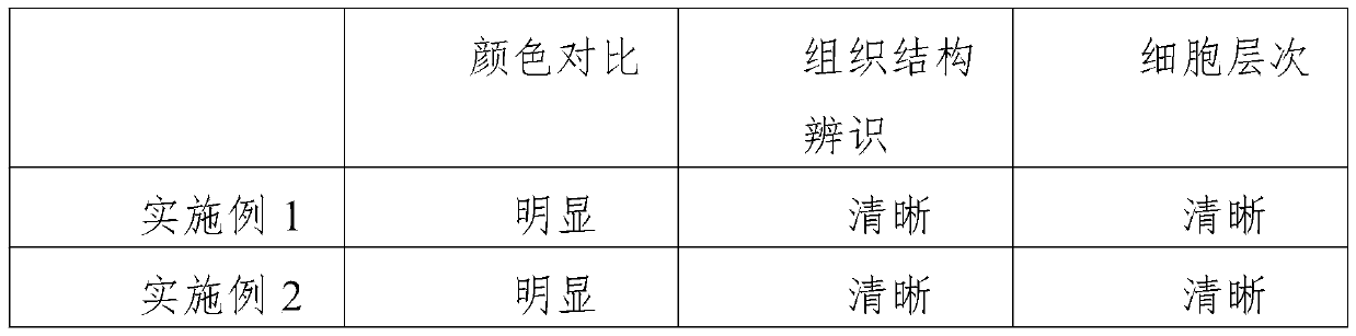
Examples
Embodiment 1
[0033] A method for processing histopathological tissue samples, including the following steps:
[0034] (1) Fixation: Fix the isolated tissue sample, and fix it with Fixative I and Fixative II in sequence;
[0035] Fixing agent I is made by mixing the following components: 2.5% glutaraldehyde 50mL, 2% paraformaldehyde 50mL; Fixing agent I is fixed at 4°C, and the fixing time is 30min;
[0036] Fixing agent Ⅱ is made by mixing the following components: 40% formaldehyde 10mL, distilled water 90mL, NaH 2 PO 4 ·H 2 O 0.5g, Na 2 HPO 4 0.5g; Fixative II is fixed at room temperature for 30 minutes.
[0037] (2) Rinse: Use running water to rinse slowly.
[0038] (3) Dehydration: gradient ethanol dehydration: 75% ethanol 30 min, 80% ethanol 30 min, 85% ethanol I 30 min, 85% ethanol II 60 min, 85% ethanol III 60 min, 95% ethanol I 40 min, 95% ethanol II 40 min, anhydrous ethanol I 30 min, no Water ethanol II 30min.
[0039] (4) Transparency: select TO transparent agent for transparency, TO trans...
Embodiment 2
[0048] A method for processing histopathological tissue samples, including the following steps:
[0049] (1) Fixation: Fix the isolated tissue sample, and fix it with fixative I and fixative II in sequence;
[0050] Fixing agent I is made by mixing the following components: 2.5% glutaraldehyde 50mL, 2% paraformaldehyde 50mL; Fixing agent I is fixed at 6°C, and the fixing time is 30min;
[0051] Fixing agent Ⅱ is made by mixing the following components: 40% formaldehyde 10mL, distilled water 90mL, NaH 2 PO 4 ·H 2 O 0.5g, Na 2 HPO 4 0.5g; Fixative II is fixed at room temperature, and the fixing time is 30min.
[0052] (2) Rinse: Use running water to rinse slowly.
[0053] (3) Dehydration: gradient ethanol dehydration: 75% ethanol 30 min, 80% ethanol 30 min, 85% ethanol I 30 min, 85% ethanol II 60 min, 85% ethanol III 60 min, 95% ethanol I 40 min, 95% ethanol II 40 min, anhydrous ethanol I 30 min, no Water ethanol II 30min.
[0054] (4) Transparency: select TO transparent agent for transpa...
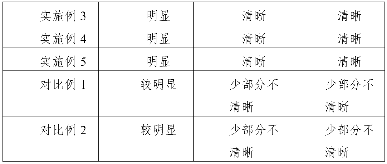
Embodiment 3
[0063] A method for processing histopathological tissue samples, including the following steps:
[0064] (1) Fixation: Fix the isolated tissue sample, and fix it with fixative I and fixative II in sequence;
[0065] Fixing agent I is made by mixing the following components: 2.5% glutaraldehyde 50mL, 2% paraformaldehyde 50mL; Fixing agent I is fixed at 6°C, and the fixing time is 30min;
[0066] Fixing agent Ⅱ is made by mixing the following components: 40% formaldehyde 10mL, distilled water 90mL, NaH 2 PO 4 ·H 2 O 0.5g, Na 2 HPO 4 0.5g; Fixative II is fixed at room temperature, and the fixing time is 30min.
[0067] (2) Rinse: Use running water to rinse slowly.
[0068] (3) Dehydration: gradient ethanol dehydration: 75% ethanol 30 min, 80% ethanol 30 min, 85% ethanol I 30 min, 85% ethanol II 60 min, 85% ethanol III 60 min, 95% ethanol I 40 min, 95% ethanol II 40 min, anhydrous ethanol I 30 min, no Water ethanol II 30min.
[0069] (4) Transparency: select TO transparent agent for transpa...
PUM
| Property | Measurement | Unit |
|---|---|---|
| melting point | aaaaa | aaaaa |
| melting point | aaaaa | aaaaa |
| melting point | aaaaa | aaaaa |
Abstract
Description
Claims
Application Information