Method for building fluorescent tumor model in nude mice based on primary cells of human cerebral glioma

A nude mouse tumor model and human glioma technology, applied to tumor/cancer cells, cells modified by introducing foreign genetic material, animal cells, etc., can solve the problems of background interference, long cycle, complicated operation, etc., and achieve Improve cycle and survival rate, stable survival period and good repeatability

Active Publication Date: 2020-05-08
武汉赛尔朗灵科技有限公司
View PDF6 Cites 6 Cited by
  • Summary
  • Abstract
  • Description
  • Claims
  • Application Information

AI Technical Summary

Problems solved by technology

[0004] 1) Because cell lines are easy to culture, economical and efficient, tumor models established with cell lines such as U251 and 9L are relatively common. For example, Chinese Patent No. CN200610000881.2 discloses a 9L that stably expresses firefly luciferase LUC The construction and application of cell lines, but because the cell lines are continuously passaged in vitro, the continuously passaged cell lines adapt to the environment of the external culture dish, gradually lose the heterogeneity of the tumor and the related signaling pathways also change, so it cannot ac

Method used


Image

  • Method for building fluorescent tumor model in nude mice based on primary cells of human cerebral glioma
  • Method for building fluorescent tumor model in nude mice based on primary cells of human cerebral glioma
  • Method for building fluorescent tumor model in nude mice based on primary cells of human cerebral glioma

Examples


Embodiment 1

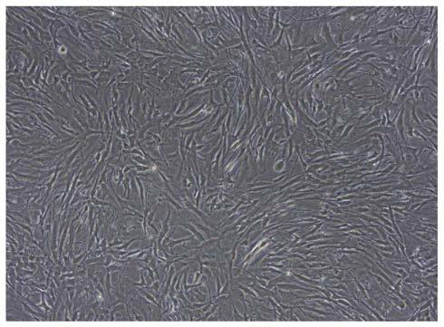
[0038] Example 1 Separation of primary human glioma cells

[0039] (1) Fresh clinical glioma resection specimens were obtained from Wuhan Union Medical College Hospital after passing the hospital ethics committee, agreeing with the patient or the patient's guardian, and signing the informed consent form. The pathological results verified that it was glioblastoma, WHO IV grade.

[0040] (2) Immediately put the resected specimen into pre-cooled sterile tissue preservation solution (containing 1000 U / mL penicillin, 1000 μg / mL streptomycin sulfate, 2.5 μg / mL amphotericin and 50 μg / mL gentamicin) Into the collection tube, and immediately put into a 4 ℃ sample transport box, transported to the laboratory within 4 hours for cell separation.

[0041] (3) Separation of primary cells: Obtain the tissue in a biological safety cabinet, rinse it once with absolute ethanol, and rinse it twice with 1×PBS (pH7.2-7.4), remove blood vessels, For fat and necrotic tissue, use dissecting scissors...
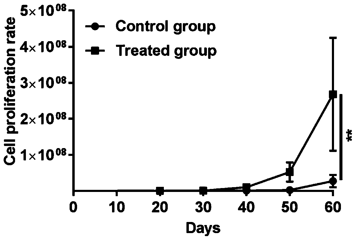
Embodiment 2

[0042] Example 2 Subculture of primary human glioma cells and optimization of subculture conditions

[0043] The isolated primary cells were divided into four groups, which were subcultured in different modified DMEM / F12 complete medium (medium A-D) conditions;

[0044] (1) Resuspend the cell pellet with modified DMEM / F12 complete medium, and store at 37°C, 5% CO 2 Cultivate under certain conditions until the cell abundance in the T25 culture flask reaches 80%, rinse the cells twice with 1×PBS (pH7.2-7.4), add 1 mL of 0.05% trypsin-EDTA to digest the monolayer cells 2- 3min.

[0045] (2) Add 2 mL of improved DMEM / F12 complete medium to stop the digestion; centrifuge at 1000 rpm for 4 minutes, remove the supernatant, collect the cell suspension, resuspend with 1 mL of improved DMEM / F12 complete medium and supplement the medium, according to the ratio of 1 to 2 , placed in a T25 culture bottle for culture, and after 20 generations of continuous passage, it is ready for use.

...

Embodiment 3

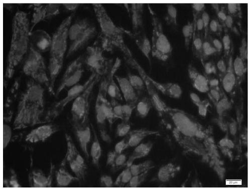
[0054] Example 3 Identification of Specific Marker Expression in Human Brain Glioma Primary Cells

[0055] (1) In the culture plate, soak the slide with the cells climbed in PBS for 3 times, each time for 3 minutes;

[0056] (2) Fix slides with 4% paraformaldehyde for 15 minutes, soak and wash slides with PBS 3 times, 3 minutes each time;

[0057] (3) Permeate with 0.5% TritonX-100 (prepared in PBS) for 20 minutes at room temperature;

[0058] (4) PBS soaked slides 3 times, 3 minutes each time, blotted the PBS with absorbent paper, added normal goat serum to the slides, and sealed at room temperature for 30 minutes;

[0059] (5) Absorb the blocking solution with absorbent paper, without washing, add a sufficient amount of diluted primary antibody to each slide and put it in a wet box, and incubate overnight at 4°C;

[0060] (6) The next day, soak the slides with PBST for 3 times, each time for 3 minutes, absorb the excess liquid on the slides with absorbent paper, add the di...


PUM


Abstract

The invention discloses a method for building a fluorescent tumor model in nude mice based on primary cells of human cerebral glioma, and belongs to the field of animal models in the medical fields. The method comprises the following steps: S1, culturing of steady-transferring cells: carrying out cell culturing by adopting an improved DMEM/F12 complete culturing medium, and massively amplifying the primary cells capable of stably expressing luciferase; S2, preparation of cell suspensions: repeatedly cleaning the cells by using the improved DMEM/F12 complete culturing medium, collecting the cell suspensions for subsequent use to ensure that the concentration of the cell suspensions is 6*10<9> cells/mL, and using the suspensions in 40 minutes after the suspensions are prepared; and S3, building of a glioma model of the nude mice: injecting the suspensions in craniums of the nude mice in situ to obtain the mice model capable of stably expressing the luciferase. The fluorescent tumor modelin nude mice based on the human cerebral glioma prepared by adopting the method has the advantages that tumor growth and pathological characteristics of the nude mice are similar to the human cerebral glioma, and growth of the tumors can be observed in real time, so that a desired research model is provided for researching glioma based on in-situ transplanting and tracing of the human cerebral glioma and researching micro-environments of the tumors.

Description

technical field [0001] The invention belongs to the field of animal models used in the medical field, and more specifically relates to a method for establishing a fluorescent nude mouse tumor model based on primary cells of human brain glioma. Background technique [0002] Glioma is a primary tumor derived from glial cells and their precursors. It is the most common and most difficult to treat malignant tumor of the central nervous system, accounting for about 50% of intracranial tumors. Due to the aggressive growth of most gliomas and the unclear boundary with the surrounding normal brain tissue, it is difficult to completely remove the tumor by surgery. Based on the fact that the biodiversity and pathogenesis of glioma cells have not yet been clarified, it is still a type of central nervous system tumor with the worst prognosis among all malignant tumors under comprehensive treatment. The average survival cycle of glioblastoma patients is only for 14.6 months. How to imp...

Claims


Application Information

Patent Timeline
IPC IPC(8): C12N5/10C12N15/867C12N7/01A01K67/027
CPCA01K67/0271A01K2207/12A01K2227/105A01K2267/0331C12N5/0693C12N7/00C12N9/0069C12N15/86C12N2740/15021C12N2740/15043
Inventor 刘红亚李俊俊李继新魏丹妮刘霞王前进王鲜
Owner 武汉赛尔朗灵科技有限公司
Why Eureka
Try Eureka