Three-dimensional biological detection system based on fluorescent quantum dots, and preparation method and application thereof

A technology of fluorescent quantum dots and three-dimensional biology, which is applied in the field of three-dimensional biological detection systems based on fluorescent quantum dots and its preparation, can solve the problems of difficulty in further improving sensitivity, difficulty in exposing antigen binding sites, and low antigen-antibody binding efficiency

Inactive Publication Date: 2020-05-29
HENAN UNIVERSITY +1
View PDF8 Cites 1 Cited by
  • Summary
  • Abstract
  • Description
  • Claims
  • Application Information

AI Technical Summary

Problems solved by technology

However, the microtiter plate has a two-dimensional structure as the base, and the captured antibodies are limited. At the same time, antibodies captured on a two-dimensional plane often have binding positions such as rear, upright, side and flat, so that the antigen binding site is difficult to expose. The combination efficiency of antigen and antibody is too low, which makes it difficult to further improve the sensitivity

Method used


Image

  • Three-dimensional biological detection system based on fluorescent quantum dots, and preparation method and application thereof
  • Three-dimensional biological detection system based on fluorescent quantum dots, and preparation method and application thereof
  • Three-dimensional biological detection system based on fluorescent quantum dots, and preparation method and application thereof

Examples


preparation example Construction

[0060] The present invention also provides a method for preparing the three-dimensional biological detection system described in the above technical solution, comprising the following steps:

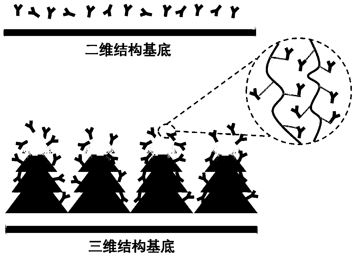
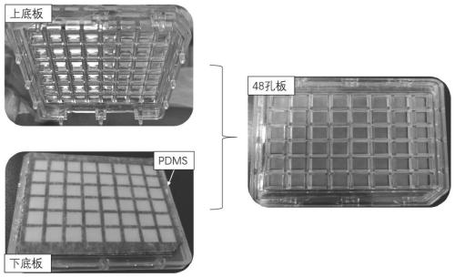
[0061] Using polydimethylsiloxane as the main body of the substrate, the surface initiates a polymerization reaction to obtain a tree-like three-dimensional structure as the substrate;

[0062] Printing antibodies on the surface of the substrate using a biochip spotter, and obtaining coated antibodies on the surface of the substrate;

[0063] Water-soluble fluorescent quantum dots are used as fluorescent probes for covalent coupling with conjugated antibodies.

[0064] In the invention, polydimethylsiloxane is used as the main body of the substrate, and the polymerization reaction is initiated on the surface to obtain a tree-like three-dimensional structure as the substrate. In the present invention, there is no special limitation on the conditions of the polymerization reaction, and me...

Embodiment 1

[0079] (1) Composition of a three-dimensional biological detection system based on fluorescent quantum dots

[0080] 1. Coating antibody: CRP monoclonal antibody, purchased from Beijing Baichuan Feihong Biotechnology Co., Ltd., the product catalog number is 7D9, and the concentration is 2.2 mg / mL. SAA monoclonal well antibody was purchased from Shanghai Unionwell Biotechnology Co., Ltd., the product catalog number is 2201, and the concentration is 5.0 mg / mL.

[0081] 2. Conjugated antibody: CRP monoclonal antibody, purchased from Hangzhou Qitai Co., Ltd., the product catalog number is MC02, and the concentration is 5 mg / mL. SAA monoclonal well antibody was purchased from Shanghai Unionwell Biotechnology Co., Ltd., the product catalog number is 2203, and the concentration is 5.1 mg / mL.

[0082] 3. Fluorescent probes: Quantum dots combined with CRP monoclonal pore antibodies to prepare QD-CRP probes; quantum dots combined with SAA monoclonal pore antibodies to prepare QD-SAA pr...

Embodiment 2

[0109] Establishment and effect evaluation of CRP detection method:

[0110] (1) Preparation of standard curve

[0111] (1) Incubation of standards: Take out the multi-well sample plate and return to room temperature. First use 1×10 in each hole -5 mol / mL PBS solution for 10 min. Dilute the CRP standard with diluent to the target concentration, add 0.1mL to each well, put it in a constant temperature culture shaker, and incubate at 37°C for 30min; shake off the reaction solution, wash with washing solution five times, and pat dry on absorbent paper.

[0112] (2) Incubation of the labeled antibody: Dilute the QD-CRP probe with diluent, 1:50 times dilution, 0.1 mL per well, put it in a constant temperature culture shaker, and incubate at 37 ° C for 30 min. Shake off the reaction solution, wash five times with washing solution, and pat dry on absorbent paper.

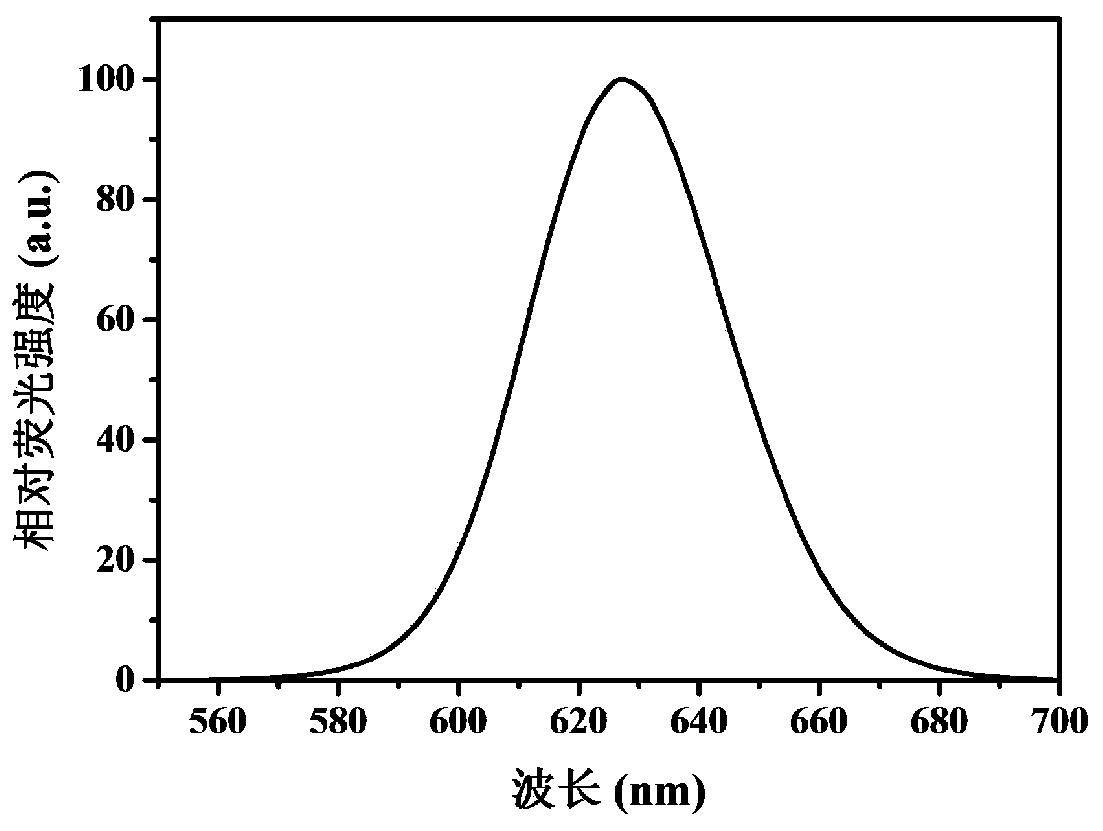
[0113] (3) Data detection: the sample is measured with a laser instrument, the excitation light wavelength is 405nm,...


PUM

PropertyMeasurementUnit
wavelengthaaaaaaaaaa
particle sizeaaaaaaaaaa
concentrationaaaaaaaaaa

Abstract

The invention provides a three-dimensional biological detection system based on fluorescent quantum dots, and a preparation method and application thereof, and belongs to the field of in-vitro diagnosis and detection. According to the three-dimensional biological detection system provided by the invention, the water-soluble fluorescent quantum dot is used as a fluorescent probe, a three-dimensional structure is combined as a detection substrate, a double-antibody sandwich method of 'coated antibody-antigen to be detected-fluorescent quantum dot labeled antibody' is utilized, and a simple, sensitive and stable novel method for detecting various markers is established. A detection signal can be amplified, the detection sensitivity can be improved, and meanwhile, the detection accuracy is ensured by good detection specificity, simplicity and convenience in operation and a stable fluorescent marker.

Description

technical field [0001] The invention relates to the technical field of in vitro diagnosis and detection, in particular to a three-dimensional biological detection system based on fluorescent quantum dots and its preparation method and application. Background technique [0002] The common liquid-phase detection method for the detection of biomarkers is enzyme-linked immunosorbent assay (ELISA), which has the advantages of rapidity, simplicity, and easy standardization of carriers, and is widely used in the quantitative determination of macromolecular antigens and small molecular antigens, as well as the Immunodiagnosis of infectious diseases, parasitic diseases and non-infectious diseases caused by microorganisms. However, the microtiter plate has a two-dimensional structure as the base, and the captured antibodies are limited. At the same time, antibodies captured on a two-dimensional plane often have binding positions such as rear, upright, side and flat, so that the antige...

Claims


Application Information

Patent Timeline
Patent Type & Authority Applications(China)
IPC IPC(8): G01N33/533G01N33/558G01N33/577
CPCG01N33/533G01N33/558G01N33/577
Inventor 李林松吕雁冰马雄明吴瑞丽申怀彬
Owner HENAN UNIVERSITY
Why Eureka
Try Eureka