Kit for detecting echinococcosis
A kit and technology for hydatid disease, applied in the field of kits, can solve the problems of low detection efficiency, missed diagnosis, difficulty in typing, misdiagnosis, etc., and achieve the effects of high detection accuracy, high detection efficiency and simple operation.
- Summary
- Abstract
- Description
- Claims
- Application Information
AI Technical Summary
Problems solved by technology
Method used
Image
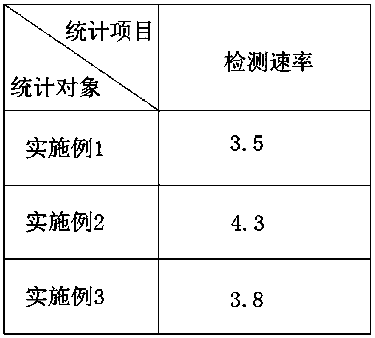
Examples
Embodiment 1
[0028] S1. Take 3 g of parasite tissue to be inspected or infected lesions of human body or animal into a test tube containing 2 ml of preservation solution. After mixing thoroughly, centrifuge at 2500 rpm for 10 minutes, extract the whole genome DNA of the sample and place it in the PCR reaction. reserve in the tube;
[0029] S2. Add 2 parts of upstream primer Eg-F, 2 parts of downstream primer Eg-R, 3 parts of dNTp mixture, 1 part of DNA polymerase, 1 part of Taq enzyme, and trimethylol into the PCR reaction tube containing the whole genome DNA of the sample in S1. 2 parts of aminomethane hydrochloric acid solution and 6 parts of sterile double distilled water, fully mixed;
[0030] S3. Place the PCR reaction tube in S2 in a PCR machine for PCR amplification, spot the amplified PCR product on a 3% agarose gel for electrophoresis, and observe the electrophoresis results, wherein the reaction condition of PCR is 94°C Pre-denaturation for 3 minutes, denaturation at 94°C for 35...
Embodiment 2
[0033] S1. Take 3 g of parasite tissue to be inspected or infected lesions of human body or animal into a test tube containing 2 ml of preservation solution. After mixing thoroughly, centrifuge at 2500 rpm for 10 minutes, extract the whole genome DNA of the sample and place it in the PCR reaction. reserve in the tube;
[0034] S2. Add 4 parts of upstream primer Eg-F, 4 parts of downstream primer Eg-R, 5 parts of dNTp mixture, 3 parts of DNA polymerase, 3 parts of Taq enzyme, and trimethylol into the PCR reaction tube containing the whole genome DNA of the sample in S1. 4 parts of aminomethane hydrochloric acid solution and 8 parts of sterile double distilled water, fully mixed;
[0035] S3. Place the PCR reaction tube in S2 in a PCR machine for PCR amplification, spot the amplified PCR product on a 3% agarose gel for electrophoresis, and observe the electrophoresis results, wherein the reaction condition of PCR is 94°C Pre-denaturation for 3 minutes, denaturation at 94°C for ...
Embodiment 3
[0038] S1. Take 3 g of parasite tissue to be inspected or infected lesions of human and animals into a test tube containing 2 ml of preservation solution. After mixing thoroughly, centrifuge at 2500 rpm for 10 minutes, extract the whole genome DNA of the sample and place it in the PCR reaction. reserve in the tube;
[0039]S2. Add 3 parts of upstream primer Eg-F, 3 parts of downstream primer Eg-R, 4 parts of dNTp mixture, 2 parts of DNA polymerase, 2 parts of Taq enzyme, and trimethylol into the PCR reaction tube containing the whole genome DNA of the sample in S1. 3 parts of aminomethane hydrochloric acid solution and 7 parts of sterile double distilled water, fully mixed;
[0040] S3. Place the PCR reaction tube in S2 in a PCR machine for PCR amplification, spot the amplified PCR product on a 3% agarose gel for electrophoresis, and observe the electrophoresis results, wherein the reaction condition of PCR is 94°C Pre-denaturation for 3 minutes, denaturation at 94°C for 35s,...
PUM
 Abstract
Description
Claims
Application Information
 - R&D
- Intellectual Property
- Life Sciences
- Materials
- Tech Scout
- Unparalleled Data Quality
- Higher Quality Content
- 60% Fewer Hallucinations
Browse by: Latest US Patents, China's latest patents, Technical Efficacy Thesaurus, Application Domain, Technology Topic, Popular Technical Reports.
© 2025 PatSnap. All rights reserved.Legal|Privacy policy|Modern Slavery Act Transparency Statement|Sitemap|About US| Contact US: help@patsnap.com

