Ultrasonic thyroid nodule benign and malignant feature visualization method based on deep learning

A thyroid nodule and deep learning technology, applied in the field of medical image processing, can solve problems such as false negative results, false positive results, inaccurate results, etc., to achieve better analysis and improve the success rate

Pending Publication Date: 2020-06-05
ZHEJIANG DE IMAGE SOLUTIONS CO LTD
View PDF0 Cites 34 Cited by
  • Summary
  • Abstract
  • Description
  • Claims
  • Application Information

AI Technical Summary

Problems solved by technology

However, needle biopsy has many limitations: needle biopsy is an invasive examination, with the risk of puncturing important blood vessels and nerves, and is not suitable for the screening of thyroid nodules in large-scale populations; when choosing a too fine needle, it may be difficult to collect Insufficient pathological specimens, too small lesions, or the operator himself did not puncture the lesion, resulting in inaccurate results; the result may be false negative or false positive, even if the result of fine needle aspiration is negative, clinically Malignancy cannot be completely ruled out
Since methods such as CAM and Grad-CAM (Gradient-based CAM) use classification-based convolutional neural networks as the basic network, early network structures such as ZeilerNet (2013) and VggNet (2014) have only a few to a dozen layers, and There is no connection between layers, and the network recognition rate is not high, resulting in unreliable visualization of benign and malignant

Method used


Image

  • Ultrasonic thyroid nodule benign and malignant feature visualization method based on deep learning
  • Ultrasonic thyroid nodule benign and malignant feature visualization method based on deep learning
  • Ultrasonic thyroid nodule benign and malignant feature visualization method based on deep learning

Examples


Embodiment Construction

[0055] The present invention will be further described in detail below in conjunction with the accompanying drawings and specific embodiments. The examples can enable those skilled in the art to understand the present invention more comprehensively, but do not limit the present invention in any way.

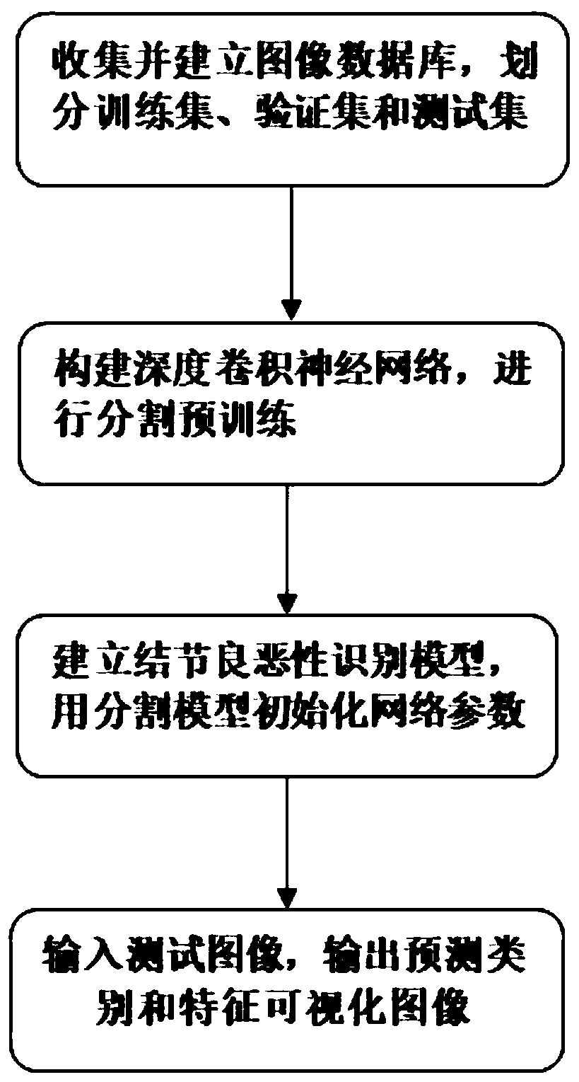
[0056] The present invention adopts a feature visualization method for identifying benign and malignant thyroid nodules based on a deep convolutional neural network, such as figure 1 As shown, the specific steps are as follows:

[0057] Process 1, collect and establish image database, divide training set, verification set and test set

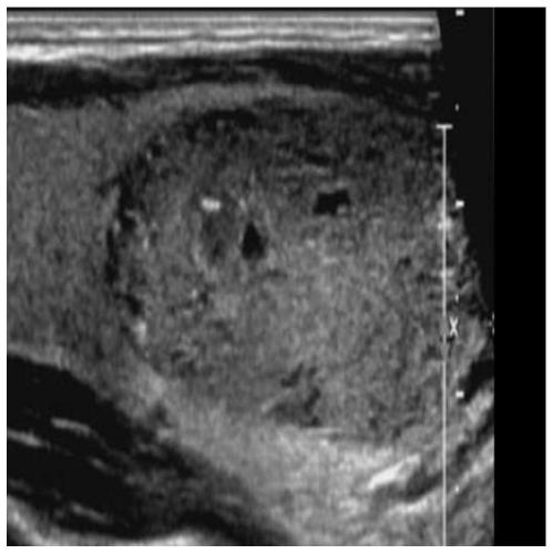
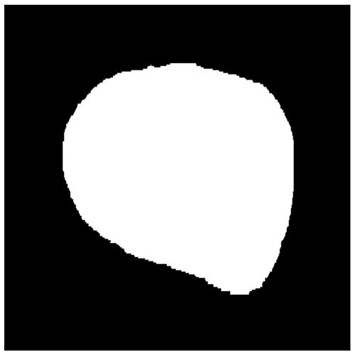
[0058] (1) Collect about 50,000 pieces of ultrasound data containing thyroid nodules, taking the case as the unit, and each case can contain multiple ordinary B-ultrasound images of different nodule sections; the cases should come from different regions and hospitals, and different ultrasound machines and operating physicians. If there are surg...


PUM


Abstract

The invention relates to a medical image processing technology, and aims to provide an ultrasonic thyroid nodule benign and malignant feature visualization method based on deep learning. The method comprises the following steps: collecting case data with both a thyroid nodule ultrasonic image and a clinical operation pathological result, distinguishing benign and malignant conditions, and markinga nodule region to generate a mask image; selecting a basic structure of a deep convolutional neural network, and performing segmentation pre-training on the mask image data of all thyroid nodules; initializing a basic network by using the model parameters, and constructing a deep convolutional neural network for identification; training and verifying in a folding intersection mode to obtain a benign and malignant recognition model; and inputting a test image, predicting an identification result by using the benign and malignant identification model, and generating a malignant feature visualization image. According to the invention, the relation between the benign and malignant probability of the nodule and the image area can be visually observed. A user can better analyze the image characteristics of the ultrasonic thyroid nodule, clinical puncture examination is further guided, and the success rate of a puncture operation is increased.

Description

technical field [0001] The present invention relates to medical image processing technology, in particular to a feature visualization method for identifying benign and malignant thyroid nodules in ultrasound images based on a deep convolutional neural network (CNN). Background technique [0002] Thyroid nodules refer to lumps in the thyroid gland, which can move up and down with the thyroid gland due to swallowing actions. It is a common clinical disease and can be caused by various etiologies. In recent years, the incidence of thyroid tumors has increased significantly, especially in some coastal cities, the incidence of malignant tumors has increased at an average annual rate of 4%, which is one of the fastest growing malignant tumors. The current incidence of thyroid nodules in China is 12.8-18.6%, of which 5-15% are malignant nodules. There are different treatment methods for different types of nodules. Surgical resection of the lesion and part or all of the thyroid tis...

Claims


Application Information

Patent Timeline
Patent Type & Authority Applications(China)
IPC IPC(8): G06T7/90G06T7/40G06T7/11G06T7/00G06T5/30G06N3/04
CPCG06T7/11G06T7/0012G06T5/30G06T7/40G06T7/90G06T2207/10132G06T2207/20132G06T2207/30096G06N3/045
Inventor 王守超
Owner ZHEJIANG DE IMAGE SOLUTIONS CO LTD
Why Eureka
Try Eureka