
Method for separating trophoblastic cells

A technology of trophoblast cells and antibodies, applied in the field of cell separation, can solve the problems of low acceptance by pregnant women, false positives, long analysis time, etc., and achieve the effect of low risk of infection and abortion, and early sampling time

Pending Publication Date: 2020-06-19
GUANGDONG HYBRIBIO BIOTECH CO LTD +2
View PDF5 Cites 7 Cited by
  • Summary
  • Abstract
  • Description
  • Claims
  • Application Information

AI Technical Summary

Problems solved by technology

This method is invasive, takes a long time to analyze, and the diagnosis time is limited to the second and third trimesters. The acceptance of pregnant women is low and it is not conducive to clinical treatment.
[0003] With the development and maturity of high-throughput sequencing technology, large-scale parallel sequencing of fetal cell-free nucleic acid with the help of high-throughput sequencing technology can accurately detect chromosomal aneuploidy. The current non-invasive prenatal diagnosis technology refers to the main It is this kind of technology, but this technology still has certain limitations, which are mainly reflected in three aspects: first, only chromosomal ploidy can be detected in current clinical applications, and the scope of inspection is extremely limited; second, it is affected by placental / maternal ploidy. Chimeric interference can easily lead to false positives. In addition, due to the calculation based on the Z value, it is difficult to completely avoid false negatives. Third, the ability to detect chromosomal deletions is insufficient, and single bases (points, insertions, deletions, etc.) cannot be tested at the same time. Fourth, there are individual differences in the content of fetal free nucleic acid

Method used


Image

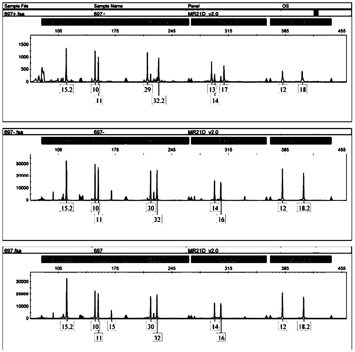
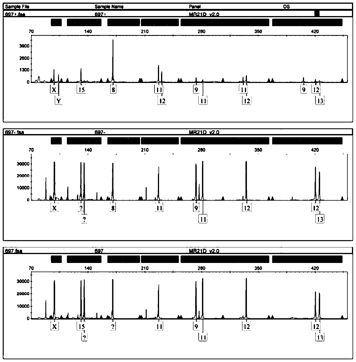
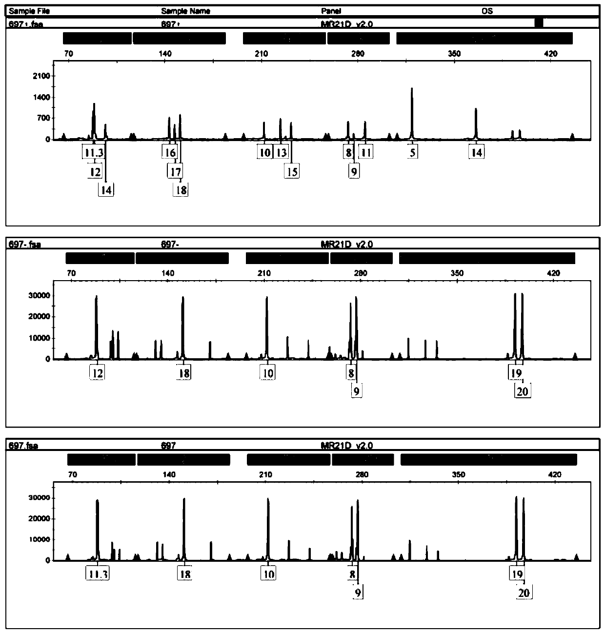
  • Method for separating trophoblastic cells
  • Method for separating trophoblastic cells
  • Method for separating trophoblastic cells

Examples


Embodiment 1

[0023] 1. Antibody selection

[0024] According to the specific antigens expressed on the surface of various types of trophoblast cells obtained by searching the literature, the antigens are shown in Table 1:

[0025] Table 1: Antigens obtained by screening

[0026]

[0027]

[0028] Cervical trophoblast cells are divided into cytotrophoblast cells, intermediate trophoblast cells, and syncytiotrophoblast cells; there is no uniform international standard for the identification of trophoblast cells, but the more recognized molecular markers are cytokeratin (cytokeratin, CK), vimentin, human chorionic gonadotropin (β-hCG), human placental lactogen (hPL), matrix metalloproteinase 9 (MMP9), etc. HLA-G molecules are expressed on embryos, extravillous trophoblast cells at the maternal-fetal interface, amniotic endothelial cells, and fetal vessel wall endothelial cells in the placenta during the entire pregnancy; trophoblast cells are cells with secretory characteristics and ca...

Embodiment 2

[0061] 1. DNA extraction from cervical exfoliated cells of pregnant women:

[0062] (1) Centrifuge 200 ul of cells from the 14th step of cell sorting (remaining cells in which trophoblast cells have been removed from cervical exfoliated cells) and sorted trophoblast cells at 12000 rpm for 3 min.

[0063] (2) Discard the supernatant, add 200ul solution P to resuspend the precipitate.

[0064] (3) Add 20ul of proteinase K and 200ul of solution L, and mix well.

[0065] (4) Incubate at 56°C for 20 minutes, and mix by inverting.

[0066] (5) Add 200 ul of absolute ethanol, mix thoroughly, transfer to an adsorption column, centrifuge at 10,000 rpm for 1 min, and discard the waste liquid in the collection tube.

[0067] (6) Add 500ul of W1, centrifuge at 10000rpm for 1min, discard the waste liquid in the collection tube.

[0068] (7) Add 500ul of W2, centrifuge at 10000rpm for 1min, discard the waste liquid in the collection tube.

[0069] (8) Add 500ul of W2, centrifuge at 1000...


PUM


Abstract

The invention provides a method for separating trophoblastic cells. The method comprises the following steps: obtaining specific antigens expressed on surfaces of various types of trophoblastic cellsaccording to searched documents, and screening the obtained specific antigens; confirming expression of the antigens in the trophoblastic cells through immunohistochemistry, and determining an antibody combination system through immunofluorescence; adding a couplant and an antibody combination into immunizing magnetic beads, and carrying out coupling; collecting a trophoblast sample from a placenta, preparing a sample cell suspension from the sample, and adding the immunizing magnetic beads with a specific antibody for incubation; and washing the immunizing magnetic beads after the incubationstep is completed, thereby obtaining separated purified trophoblastic cells of the placenta. Compared with traditional amniocentesis and chorionic villus sampling, the method has a noninvasive advantage, the material drawing time is relatively early, the risk of infection and abortion is low, and detection results have similar reliability.

Description

technical field [0001] The invention relates to the technical field of cell separation, in particular to a kit for separating trophoblast cells and an application thereof. Background technique [0002] Prenatal diagnosis is one of the important means to reduce birth defects and improve population quality. At present, the main method of clinical prenatal diagnosis is to obtain fetal samples through amniocentesis or umbilical vein puncture for karyotype analysis. This method is invasive, takes a long time to analyze, and the diagnosis time is limited to the second and third trimesters. The acceptance of pregnant women is low and it is not conducive to clinical treatment. [0003] With the development and maturity of high-throughput sequencing technology, large-scale parallel sequencing of fetal cell-free nucleic acid with the help of high-throughput sequencing technology can accurately detect chromosomal aneuploidy. The current non-invasive prenatal diagnosis technology refers...

Claims


Application Information

Patent Timeline
Patent Type & Authority Applications(China)
IPC IPC(8): C12N5/073C12Q1/6888
CPCC12N5/0605C12Q1/6888
Inventor 郑焱马佳艳丰晓威薛晋杰陈建勇
Owner GUANGDONG HYBRIBIO BIOTECH CO LTD
Learn More