
Wide-field color light slice microscopic imaging method based on deep learning

A technology of microscopic imaging and deep learning, applied in neural learning methods, image enhancement, image analysis, etc., can solve problems such as complex defocus information, poor resolution, and large data collection required for SIM-OS imaging, and achieve Effects of high temporal resolution, increased imaging throughput, and avoided risk of phototoxic contamination

Pending Publication Date: 2020-07-17
MGI TECH CO LTD
View PDF3 Cites 3 Cited by
  • Summary
  • Abstract
  • Description
  • Claims
  • Application Information

AI Technical Summary

Problems solved by technology

[0006] The purpose of the present invention is to combine deep learning, wide-field microscopic imaging, data fusion and other technologies to provide a wide-field color light slice microscopic imaging method based on deep learning, that is, FC-WFM-deep, to overcome wide-field microscopic imaging. In imaging, the defocus information is complex, the resolution is not good, and SIM-OS imaging requires a large amount of data collection, etc.

Method used


Image

  • Wide-field color light slice microscopic imaging method based on deep learning
  • Wide-field color light slice microscopic imaging method based on deep learning
  • Wide-field color light slice microscopic imaging method based on deep learning

Examples


Embodiment Construction

[0058] The present invention will be described in detail below in conjunction with the accompanying drawings and embodiments.

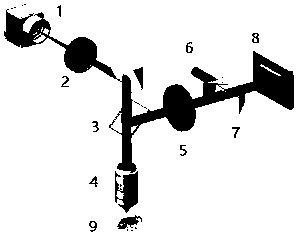
[0059] Please refer to figure 1 As shown, the present invention exemplifies a full-color microscopy imaging system that performs raw WFM and FC-SIM imaging using a collimated high-power white LED (SILIS-3C, Thorlabs, Inc.) as the illumination source. Then, the LED light enters a total internal reflection prism (TIR prism) and is reflected to a DMD (V7000, ViALUX GmbH, Germany). Afterwards, the modulated light passed through an optical projection system, including an achromatic collimator lens, a beam splitter, and an objective lens (20x objective lens, NA = 0.45, Nikon Inc., Japan), and projected a sinusoidal fringe pattern onto the sample. The sample was mounted on an x-y-z motorized translation table (Ataucube, Germany). A color camera (80FPS, 2048 × 2048 pixels, IDS GMH, Germany) was used to capture 2D widefield images of the scans.

[0060] Ple...


PUM


Abstract

The invention belongs to the technical field of optical microscopy imaging, provides a wide-field color light slice microscopy imaging method based on deep learning, and solves the problems that defocusing information is complex, the resolution ratio is poor, SIM-OS imaging needs large-data-volume acquisition and the like in wide-field microscopy imaging. Unique high resolution and panchromatic property of full-color structured light illumination are fully utilized, and a single-width wide-field microscopic imaging result can be used for training. The method is popularized to a wide-field microscopic imaging experiment; according to the method, a light slice component with a wide scene depth and a full-color high-quality image can be directly obtained from a wide-field image, and meanwhile, the method has three-dimensional reconstruction and data analysis capabilities equivalent to those of full-color structured light illumination imaging in spatial resolution and dimension; in addition, the required data volume is sharply reduced compared with that of full-color structured light illumination; according to the method, under the condition that details are not lost, the imaging throughput of an imaging system is remarkably improved by extracting a wide-scene-depth optical slice result and reducing data acquisition, and meanwhile, the phototoxicity pollution risk of the system isreduced.

Description

technical field [0001] The invention belongs to the technical field of optical microscopic imaging, and specifically relates to deep learning technology, combined with full-color wide-field microscopic imaging, proposes a deep learning-based wide-field color light slice microscopic imaging with low data acquisition and high space-time resolution method. Background technique [0002] Wide-field microscopy is a basic sample imaging method in microscopy, which exposes the entire sample of interest to a light source through an observer, camera, or computer monitor to obtain an observation image. Compared with confocal microscopy or electron microscopy, wide-field microscopy (WFM), as a low-cost, fast imaging, and low photobleaching imaging mode, is usually the imaging tool of choice for biologists to conduct research. However, WFM not only collects the light emitted by the target in the focal plane, but also superimposes all the light emitted by the illuminated layer of the sam...

Claims


Application Information

Patent Timeline
IPC IPC(8): G06T17/00G06T7/593G06T7/90G06K9/62G06N3/04G06N3/08G01N21/84
CPCG06T17/00G06T7/596G06T7/90G06N3/08G01N21/84G06T2207/10061G06N3/045G06F18/25
Inventor 柏晨姚保利但旦千佳党诗沛
Owner MGI TECH CO LTD
Learn More