
Brain MRI image segmentation method based on self-organizing mapping network for medical treatment and MRI equipment

A technology of self-organizing mapping and image segmentation, applied in image analysis, image enhancement, image data processing, etc., can solve problems such as lack of convergence criteria, lack of automation, and influence

Inactive Publication Date: 2020-10-09
山东凯鑫宏业生物科技有限公司
View PDF0 Cites 1 Cited by
  • Summary
  • Abstract
  • Description
  • Claims
  • Application Information

AI Technical Summary

Problems solved by technology

[0003] In the existing segmentation method, the segmentation method of manually marking the boundary requires the operator to draw the edge of the target object to be segmented in each frame of the image. The whole process requires manual operation and lacks automation, and the detection results are subject to manipulation. The subjective factors of personnel have a greater influence
The segmentation method based on region growth must set seed points, the algorithm execution speed is slow, and there is a lack of general convergence criteria, and the selection of seed points and convergence criteria has a great impact on the results
The segmentation method based on the traditional active contour model can achieve 3D target object segmentation with less manual intervention, but this method is weak in processing complex topological structures, and performance indicators such as segmentation effect and segmentation accuracy are difficult to meet the needs of clinical applications.

Method used

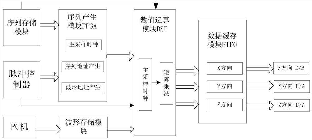
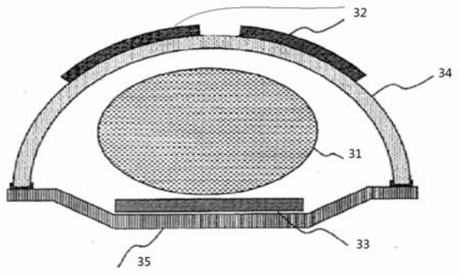

Image

  • Brain MRI image segmentation method based on self-organizing mapping network for medical treatment and MRI equipment
  • Brain MRI image segmentation method based on self-organizing mapping network for medical treatment and MRI equipment
  • Brain MRI image segmentation method based on self-organizing mapping network for medical treatment and MRI equipment

Examples


Embodiment Construction

[0059] The present invention will be further described below in conjunction with the accompanying drawings.

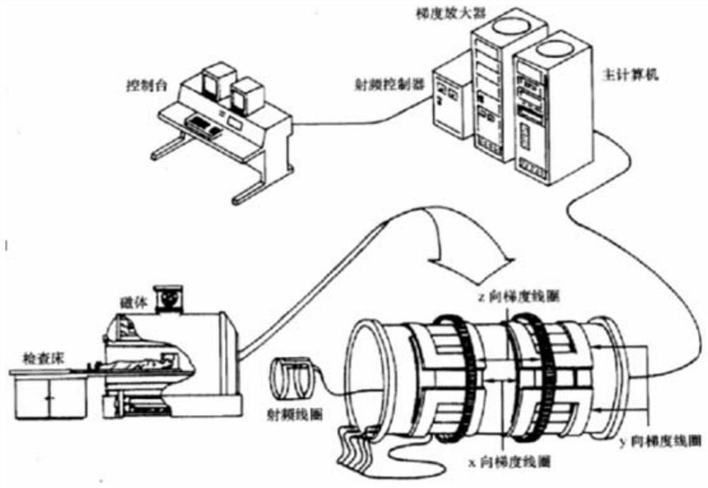
[0060] In an embodiment of the present invention, a magnetic resonance imaging device based on multiple receiving coils includes: a main magnet system, a gradient magnetic field system, a radio frequency system, and an operation and image processing system.

[0061] 1. Main magnet system

[0062] The double-column main magnet used in this application mainly includes components such as a magnet, a yoke, a pole shoe and a frame. The magnet is designed as a left and right cylinder, which provides magnetic energy and generates an imaging static magnetic field. The material is NdFeB; the frame is used to support the magnet structure; , to ensure that the magnetic field strength and distribution in the magnetic field work area meet the predetermined requirements, and reduce magnetic flux leakage. The material is usually steel. In this main magnet, the surfaces of the upper...


PUM


Abstract

The invention relates to a brain MRI image segmentation method based on a self-organizing mapping network and MRI equipment. The method comprises the following steps: step (1), performing feature extraction on an input fused brain MRI image; (2), designing a multi-layer neural network according to the characteristics of the brain tumor image, and training the neural network by selecting a brain MRI image which is calibrated in advance; (3), sending the whole image to the trained network through each pixel, and for the feature vector of each pixel, generating a winning neuron on the first layeraccording to the minimum distance standard so as to specify a corresponding object by using the pixel; and step (4), during a merging process, in order to suppress wrong classification, requiring a merging clustering process to connect neurons belonging to normal classification after segmentation so as to segment a tumor region effectively and obtain a target image. The method can effectively jump out of local optimum, and is high in brain tumor MRI image segmentation precision and smooth in edge.

Description

technical field [0001] The invention relates to the technical field of MRI image acquisition and medical image processing, in particular to a brain tumor segmentation method in an MRI brain image and MRI equipment. Background technique [0002] Magnetic resonance imaging (MRI) has the advantages of low radiation, high sensitivity of soft tissue imaging, multi-directional imaging, and multiple imaging methods, and has been widely used. At present, MRI has become one of the important means for medical workers to study the brain. Its main advantages are: (1) it can clearly show soft tissue, anatomical structure and lesion shape; (2) various parameters can be used to adjust the imaging results, and at the same time obtain rich Diagnostic information; (3) Any section can be imaged, and images of parts that are difficult to access by other imaging techniques can be obtained; (4) There is no ionizing radiation damage to the human body, and it is safe and non-invasive to the human b...

Claims


Application Information

Patent Timeline
IPC IPC(8): G06T7/11G06T5/00G06T5/50G06T7/00G06K9/62G06N3/04G06N3/08G06F17/16
CPCG06T7/11G06T5/50G06T7/0012G06N3/08G06F17/16G06T2207/10088G06T2207/30016G06T2207/30096G06T2207/20221G06N3/045G06F18/23G06F18/22G06T5/70
Inventor 冯叶
Owner 山东凯鑫宏业生物科技有限公司
Learn More