Immune cell organoid co-cultures

A technology of co-culture and immune cells, applied in the field of co-culture of immune cell organoids, can solve the problems of immune cell co-culture cancer treatment plan, no report of human organoid co-culture progress cancer application and other problems

Pending Publication Date: 2020-11-24
KONINK NEDERLANDSE AKADE VAN WETENSCHAPPEN +1
View PDF2 Cites 9 Cited by
  • Summary
  • Abstract
  • Description
  • Claims
  • Application Information

AI Technical Summary

Problems solved by technology

[0006] Previous attempts have demonstrated co-cultures of murine intraepithelial lymphocytes (IEL) with murine intestinal epithelial organoids with the aim of understanding the spatiotemporal behavior of IELs and intestinal epithelial cells - Nozaki et al. (J Gastroenterol. 2016 3 51(3):206-13) and Rogoz et al. (J Immunol Methods. 2015 Jun; 421:89-95) – but no progress has been reported in the development of co-cultures of human organoids and in the study and treatment of cancer Applications
So-called 'tumor-like' have been prepared from samples derived from colorectal cancer patients (Drost et al., Nature. 2015 May 7;521(7550):43-7; van de Wetering et al., Cell. 2015 May 7;161(4):933-45), but not co-cultured with immune cells to study cancer treatment options

Method used


Image

  • Immune cell organoid co-cultures
  • Immune cell organoid co-cultures
  • Immune cell organoid co-cultures

Examples


Embodiment 1

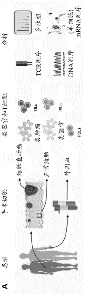
[0273] Example 1. Collection of normal colon and colorectal cancer biopsies from a hospital.

[0274] This example shows the isolation of cell samples, which are used in subsequent examples to prepare organoid, tumor and immune cell samples.

[0275] Biopsies of normal colonic mucosa and tumor tissue were obtained from the resected colon and / or rectum of patients with colorectal cancer. Peripheral blood was also drawn during surgery.

[0276] Specifically, biopsy tissues from human colorectal cancer tissues as well as normal (adult) human colonic mucosal epithelium were collected in 50 mL standard tubes containing ice-cold 10-15 mL of high-grade DMEM / F12 medium completely With penicillin / streptomycin (from 100× stock solution of 10,000 U / mL penicillin and 10K μM / mL streptomycin), HEPES (from 100× stock solution of 1M), GlutaMAX (from 100× stock solution; all Gibco TM ) and the Rho kinase inhibitor Y-27632 (Sigma-Aldrich). Biopsies were kept on ice and processed immediately,...

Embodiment 2

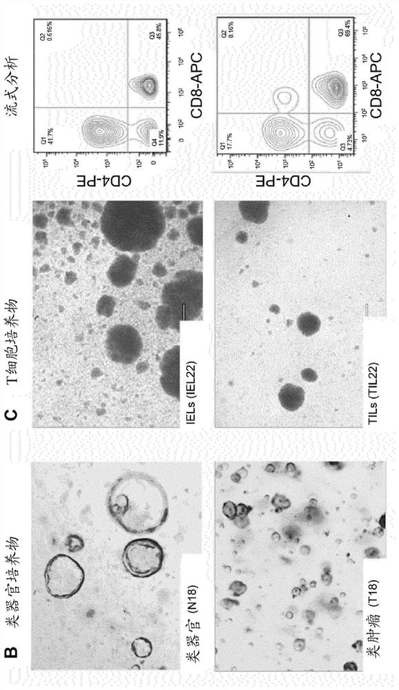
[0278] Example 2. Isolation of crypts from normal colon tissue and derivation of normal colon organoids; isolation of intraepithelial T cells from normal colon tissue for T cell culture.

[0279] This example shows the processing of normal colon samples for the development of organoid cultures and for the isolation of immune cells from normal colon samples.

[0280] Normal colonic mucosa was treated with EDTA to release crypts for deriving normal colonic organoids, and then further digested to prepare single-cell suspensions containing intraepithelial lymphocytes (IELs) for T cell culture.

[0281] Isolation of crypts and derivation of normal colon organoids from normal colon tissue.

[0282] Remove the muscle layer and fat under a dissecting microscope using surgical scissors and forceps. Cut the cleaned tissue into thin strips of approximately 1-2 mm. One strip was fixed in 4% formaldehyde (Sigma-Aldrich) for histological analysis, and one strip was snap frozen (in dry ice...

Embodiment 3

[0286] Example 3. Digestion of colorectal cancer tissue for tumor organoid and T cell culture; derivation of colorectal cancer tumoroids.

[0287] This example shows the processing of cancerous colon samples for the development of tumor-like cultures, and the isolation of immune cells from cancerous colon samples.

[0288] Tumor tissue was digested to prepare single-cell suspensions containing epithelial tumor cells, tumor-infiltrating lymphocytes (TILs) for deriving tumoroids, and for T-cell culture.

[0289] Digestion of colorectal cancer tissue for tumor and T cell culture.

[0290] Tumor biopsies were cut into thin strips of approximately 1-2 mm. One strip was fixed in 4% formaldehyde for histological analysis, and each strip was snap frozen (in dry ice or liquid nitrogen) and stored at -80°C for genetic and / or protein analysis. The remaining strips were further cut using forceps until the tumor mass appeared viscous. Tumor masses were incubated in 10 mL of complete hig...


PUM


Abstract

The present invention provides co-cultures of organoids and immune cells, and methods of using these to identify agents for treating diseases.

Description

[0001] All documents cited herein are incorporated by reference in their entirety. technical field [0002] The present invention relates to organoid co-cultures and their use in disease research. Background technique [0003] While the in vitro systems used to conduct such research remain fundamental, clinical studies of physiologically based diseases such as cancer and immune disorders remain a cornerstone of medical progress. Likewise, modern regimens for treating such diseases often involve rigorous testing systems during development to ensure the regimen's efficacy and safety. Although recent advances in these fields have increased the efficacy of research and therapeutic testing systems, improvements in the efficiency, accuracy, and cost-effectiveness of the systems are needed. An ideal testing system would precisely and accurately replicate the physiology of a patient or patient population at the biochemical, cellular, tissue, organ, and organismal level, eliminating...

Claims


Application Information

Patent Timeline
Patent Type & Authority Applications(China)
IPC IPC(8): G01N33/50C12N5/09
CPCC12N5/0693G01N33/5008G01N2500/10C12N2502/11C12N2503/02C12N2503/04G06V20/698C12Q1/6869G01N33/5088C12Q2600/106C12N5/0634C12N2501/2302C12N2501/2307C12N2501/2315G01N33/5011G01N33/5082C12N2502/30
Inventor 凯·克里施玛尔约坦·埃拉扎尔·巴-艾普生约翰尼斯·卡洛斯·克莱维尔斯西尔维娅·费南德斯-波吉罗伯特·格哈达斯·雅各布·弗里斯
Owner KONINK NEDERLANDSE AKADE VAN WETENSCHAPPEN
Why Eureka
Try Eureka