Blood vessel three-dimensional modeling method, device and system with narrow lesion interval

A technology of three-dimensional modeling and blood vessels, which is applied in the fields of three-dimensional modeling of blood vessels, coronary artery analysis systems and computer storage media, and can solve the problem that two-dimensional coronary angiography images cannot accurately determine the stenotic lesion interval, etc.

Pending Publication Date: 2020-12-25
SUZHOU RAINMED MEDICAL TECH CO LTD
View PDF0 Cites 7 Cited by
  • Summary
  • Abstract
  • Description
  • Claims
  • Application Information

AI Technical Summary

Problems solved by technology

[0006] The present invention provides a three-dimensional modeling method, device and system for blood vessels with narrow lesion intervals, so as

Method used


Image

  • Blood vessel three-dimensional modeling method, device and system with narrow lesion interval
  • Blood vessel three-dimensional modeling method, device and system with narrow lesion interval
  • Blood vessel three-dimensional modeling method, device and system with narrow lesion interval

Examples


Embodiment 1

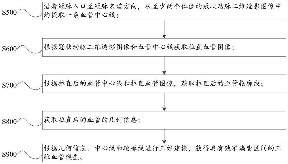
[0111] Such as figure 1 As shown, in order to solve the above problems, the present application provides a three-dimensional modeling method of blood vessels with narrow lesion intervals, including:

[0112] S500, extracting a blood vessel centerline from coronary two-dimensional angiography images of at least two body positions along the direction from the coronary artery entrance to the end of the coronary artery;

[0113] S600, acquiring a straightened blood vessel image according to the two-dimensional coronary angiography image and the center line of the blood vessel;

[0114] S700. Obtain the straightened vessel contour line according to the straightened vessel center line and the straightened vessel image;

[0115] S800, acquiring geometric information of the straightened blood vessel;

[0116] S900, performing three-dimensional modeling according to the geometric information, centerline and contour line, to obtain a three-dimensional blood vessel model with a narrow ...

Embodiment 2

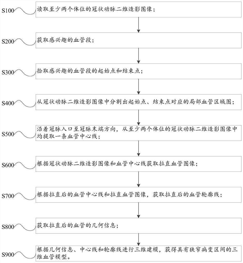
[0118] Such as figure 2 As shown, in order to solve the above problems, the present application provides a three-dimensional modeling method of blood vessels with narrow lesion intervals, including:

[0119] S100, reading coronary two-dimensional angiography images of at least two body positions; preferably, the angle between the two body positions is greater than or equal to 30 degrees;

[0120] S200, acquiring a blood vessel segment of interest;

[0121] S300, pick up the starting point and the ending point of the vessel segment of interest;

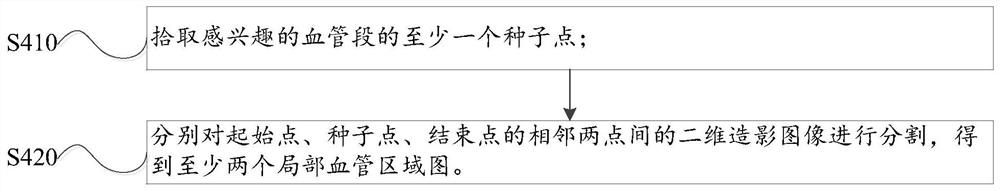
[0122] S400, segment the local blood vessel region map corresponding to the starting point and the ending point from the two-dimensional coronary angiography image, such as image 3 shown, including:

[0123] S410, picking at least one seed point of the vessel segment of interest;

[0124] S420. Segment the two-dimensional contrast images between two adjacent points of the start point, the seed point, and the end point, respective...

Embodiment 3

[0173] Such as Figure 14 As shown, the present application provides a three-dimensional modeling device for blood vessels with narrow lesion intervals, including: a central line extraction unit 100, a straightening unit 200, a contour line unit 300, a geometric information unit 400 and a three-dimensional modeling unit connected in sequence 500; the straightening unit 200 is connected to the geometric information unit 400, and the three-dimensional modeling unit 500 is connected to the straightening unit 200 and the contour line unit 300; the centerline extraction unit 100 is used to follow the direction from the coronary artery entrance to the end of the coronary artery, from at least A blood vessel centerline is extracted from the two-dimensional coronary angiography images of the two body positions; the straightening unit 200 is used to receive the blood vessel centerline sent by the centerline extraction unit 100, and obtain the straightening according to the two-dimension...


PUM


Abstract

The invention provides a method, a device and a system for acquiring a vascular stenosis lesion interval and performing three-dimensional synthesis. A blood vessel center line is extracted from coronary artery two-dimensional contrast images of the at least two body positions in the direction from the coronary artery inlet to the coronary artery tail end; A straightened blood vessel image is acquired according to the coronary artery two-dimensional contrast image and a blood vessel center line; a straightened blood vessel contour line is acquired according to the straightened blood vessel center line and the straightened blood vessel image; geometric information of the straightened blood vessel is acquired. Three-dimensional modeling is carried out according to the geometric information, the center line and the contour line to obtain a three-dimensional blood vessel model with a narrow lesion interval. The stenosis lesion interval is stereoscopically displayed to a patient and a doctorin a three-dimensional blood vessel model mode, the stenosis lesion interval is more vivid, all angles of the stenosis lesion can be displayed, the influence of the shooting angle of the two-dimensional radiography image on the judgment result of coronary artery stenosis is reduced, and the stenosis lesion area of the blood vessel can be accurately judged.

Description

technical field [0001] The invention relates to the technical field of coronary artery medicine, in particular to a three-dimensional modeling method and device for blood vessels with narrow lesion intervals, a coronary artery analysis system and a computer storage medium. Background technique [0002] The deposition of lipids and sugars in human blood on the blood vessel wall will form plaques on the blood vessel wall, which in turn will lead to vascular stenosis; especially the vascular stenosis near the coronary artery of the heart will lead to insufficient blood supply to the myocardium and induce coronary heart disease , angina pectoris and other diseases pose a serious threat to human health. According to statistics, there are about 11 million patients with coronary heart disease in my country, and the number of patients treated with cardiovascular interventional surgery is increasing by more than 10% every year. [0003] Conventional medical detection methods such as...

Claims


Application Information

Patent Timeline
IPC IPC(8): G06T17/00G06T7/13
CPCG06T17/00G06T7/13G06T2207/30101
Inventor 王之元刘广志曹文斌吴心娱
Owner SUZHOU RAINMED MEDICAL TECH CO LTD
Why Eureka
Try Eureka