Aortic dissection evaluation method and device, equipment and storage medium

A technology for aortic dissection and aorta, which is applied in the field of image processing and can solve the problems of inability to intuitively reflect vascular lesions and single information.

Pending Publication Date: 2020-12-29
SHANGHAI UNITED IMAGING HEALTHCARE
View PDF6 Cites 2 Cited by
  • Summary
  • Abstract
  • Description
  • Claims
  • Application Information

AI Technical Summary

Problems solved by technology

Doctors analyze and process the lesion by focusing on vascular morphological indicators (the degree of vascular stenosis, the degree of false lumen expansion, the size of the tear site, etc.).

Method used


Image

  • Aortic dissection evaluation method and device, equipment and storage medium
  • Aortic dissection evaluation method and device, equipment and storage medium
  • Aortic dissection evaluation method and device, equipment and storage medium

Examples


Embodiment 1

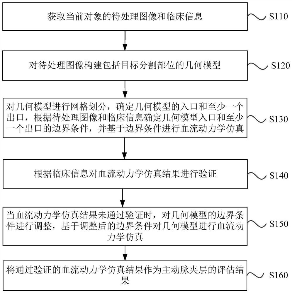
[0030] figure 1 It is a schematic flowchart of a method for assessing aortic dissection provided by Embodiment 1 of the present invention. This embodiment can be applied to perform hemodynamic simulation on the geometric model constructed from images to be processed, and determine the main aortic dissection according to the hemodynamic simulation results. In the case of the evaluation result of arterial dissection, the method can be executed by an aortic dissection evaluation device, wherein the system can be implemented by software and / or hardware, and is generally integrated in an aortic dissection evaluation device. For details, see figure 1 As shown, the method may include the following steps:

[0031] S110. Acquire the image to be processed and clinical information of the current object.

[0032] Wherein, the current object may be the examinee or the scanned part of the examinee, for example, the chest or head of the examinee. The image to be processed may be a magneti...

Embodiment 2

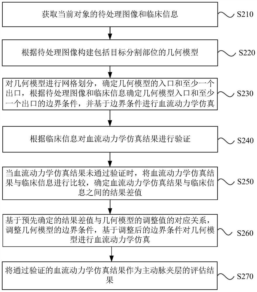
[0050] image 3 It is a schematic flowchart of a method for assessing aortic dissection provided in Embodiment 2 of the present invention. The technical solution of this embodiment is refined on the basis of the above embodiments. Optionally, when the hemodynamic simulation results fail to pass the verification, the boundary conditions of the geometric model are adjusted, including : Comparing the hemodynamic simulation result with the clinical information, determining the result difference between the hemodynamic simulation result and the clinical information; based on the pre-determined result difference and the adjustment value of the geometric model Adjust the boundary conditions of the geometric model, wherein the corresponding relationship is obtained by training the original parameter adjustment model according to the sample simulation results, sample clinical information and sample adjustment values. For the parts not described in detail in the method embodiment, plea...

Embodiment 3

[0065] Figure 4 It is a schematic structural diagram of an evaluation device for aortic dissection provided in Embodiment 3 of the present invention. see Figure 4 As shown, the device includes: a data acquisition module 310 , a geometric model construction module 320 , a boundary condition determination module 330 , a hemodynamic simulation module 340 , a verification module 350 , a boundary condition adjustment module 360 ​​and an evaluation result determination module 370 .

[0066] Wherein, the data acquisition module 310 is used to acquire images to be processed and clinical information of the current object;

[0067] A geometric model construction module 320, configured to construct a geometric model including a target segmentation site according to the image to be processed, wherein the target segmentation site includes the aorta and at least some aortic branches;

[0068] The boundary condition determination module 330 is configured to mesh the geometric model, dete...


PUM


Abstract

The embodiment of the invention discloses an aortic dissection evaluation method anddevice, equipment and a storage medium. The method comprises acquiring a to-be-processed image and clinical information of a current object, and constructing a geometric model comprising a target segmentation part according to the to-be-processed image; meshing the geometric model, determining an inlet and at leastone outlet of the geometric model, determining boundary conditions of the inlet and at least one outlet of the geometric model according to the to-be-processed image and the clinical information, andperforming hemodynamic simulation based on the boundary conditions; verifying the hemodynamic simulation result according to the clinical information; when the hemodynamic simulation result does notpass the verification, adjusting the boundary conditions of the geometric model, and performing hemodynamic simulation on the geometric model based on the adjusted boundary conditions; and taking theverified hemodynamic simulation result as an evaluation result of the aortic dissection. The aortic dissection is evaluated from the perspective of hemodynamics, and the accuracy of an aortic dissection evaluation result is improved.

Description

technical field [0001] Embodiments of the present invention relate to image processing technology, and in particular to a method, device, equipment and storage medium for evaluating aortic dissection. Background technique [0002] Aortic dissection (AD for short) usually means that the blood in the aortic lumen enters the aortic media-adventitia or the junction of media and adventitia through the intima tear on the aortic wall, tearing the aortic wall into two layers , a condition in which true lumen and false lumen are formed and extend along the longitudinal axis of the aorta, which can lead to aortic rupture in a short period of time and cause death of the patient, or the true lumen of the dissection is compressed by the false lumen to cause stenosis or even occlusion, and the blood is supplied by the true lumen Ischemic changes in vital organs can cause serious complications. [0003] In the prior art, blood vessel analysis software is generally used for blood vessel an...

Claims


Application Information

Patent Timeline
Patent Type & Authority Applications(China)
IPC IPC(8): G06T7/00G06T7/10G06T17/20G16H50/50G06N3/04
CPCG06T7/0012G06T7/10G06T17/205G16H50/50G06T2207/30101G06T2207/20081G06T2207/20084G06T2210/24G06T2210/41G06N3/044G06N3/045
Inventor 郭健吴柯
Owner SHANGHAI UNITED IMAGING HEALTHCARE
Why Eureka
Try Eureka