DNA framework-coated G-quadruplex structure as well as preparation method and application thereof

A framework structure and DNA strand technology, applied in the field of DNA framework-wrapped G-quadruplex structure and its preparation, can solve the problems of high background signal, poor permeability and the like, and achieve low cost, simple preparation method and high cell permeability effect on the ability to hybridize to the target

Pending Publication Date: 2021-01-15
NANTONG UNIVERSITY
View PDF0 Cites 0 Cited by
  • Summary
  • Abstract
  • Description
  • Claims
  • Application Information

AI Technical Summary

Problems solved by technology

Despite the multiple advantages of G-quadruplexes, their use in extracellular biosensing and detection in living cells remains a challenge due to poor cell permeability and high background signal of G-quadruplexes

Method used


Image

  • DNA framework-coated G-quadruplex structure as well as preparation method and application thereof
  • DNA framework-coated G-quadruplex structure as well as preparation method and application thereof
  • DNA framework-coated G-quadruplex structure as well as preparation method and application thereof

Examples


Embodiment 1

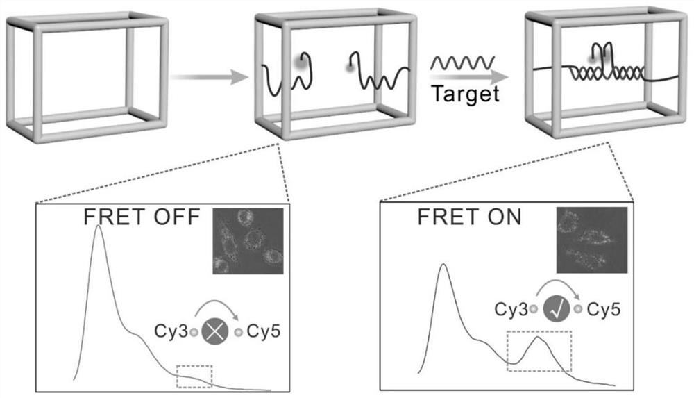
[0029] The 8 sequences used to assemble the DNA framework were first quantified to 100 μM, wherein the 8 sequences included the 6 DNA sequences C1-C6 required for the assembly of the framework structure, and the Cy3 sequence C7a required to form the G-quadruplex probe and Cy5 sequence C7b, the linear G-quadruplex probe targets miR-122a, as shown in Table 1 below:

[0030] Table 1 Assembled G-quadruplex frame-related DNA sequences

[0031]

[0032]

[0033] Then mix evenly in 100 μL of 1×TAMg buffer at a final concentration of 0.6 μM, put the centrifuge tube into a PCR machine and cool down from 95 °C to 4 °C, and the cooling time lasts for 3.5 hours, and the DNA frame-wrapped G- The quadruplex structure, such as figure 1 shown.

Embodiment 2

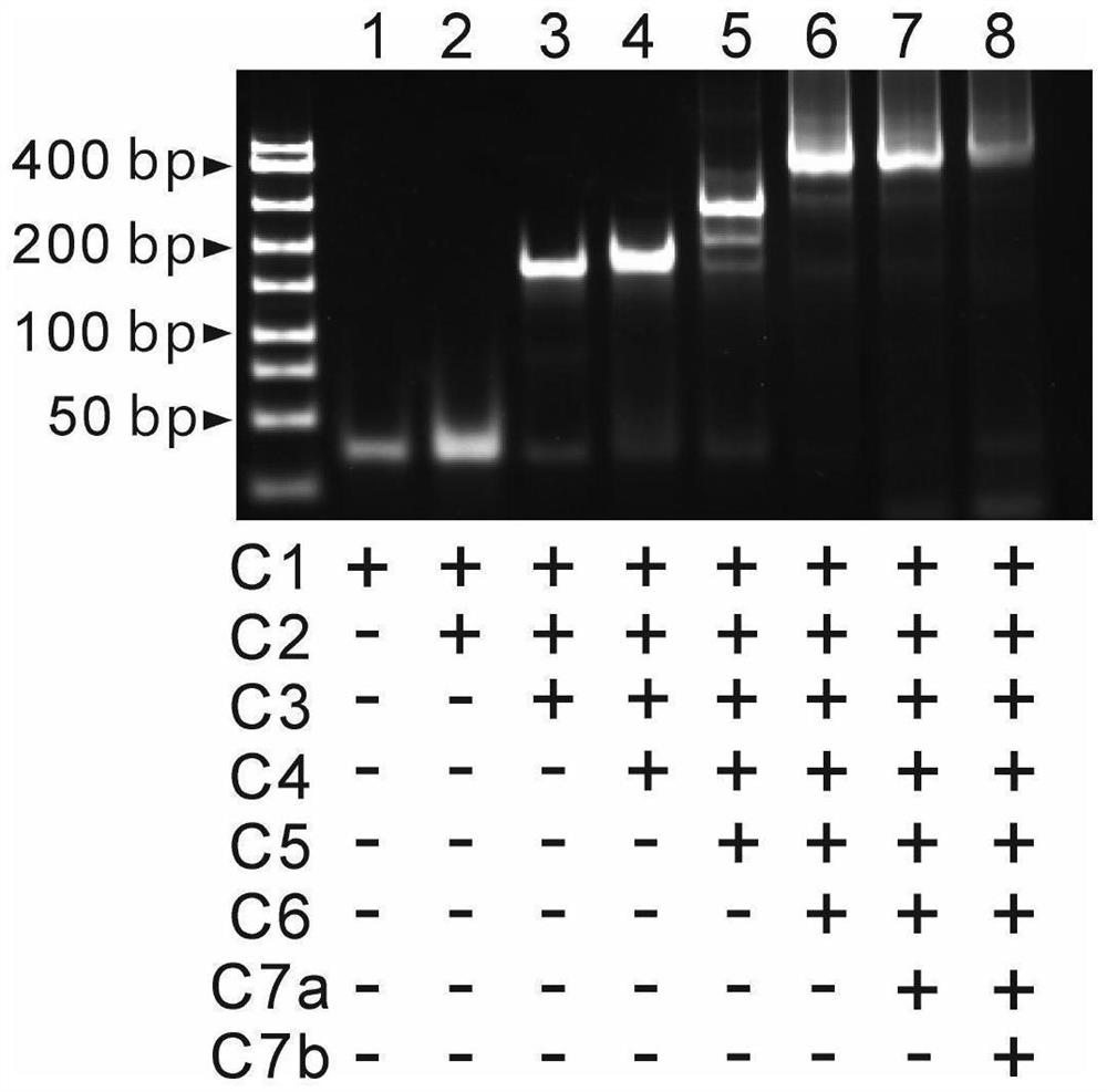
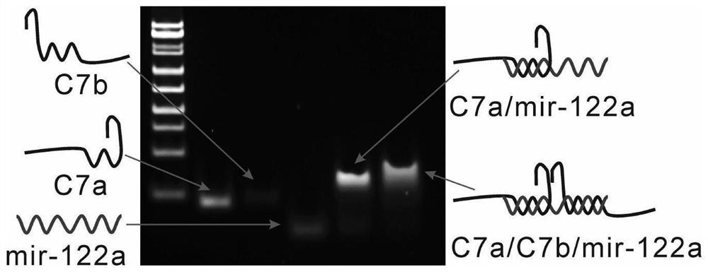
[0035] Prepare 6% PAGE gel with 5×TBE1.2mL, 30% Arc.Bis.1.2mL, three-distilled water 3.6mL, ammonium persulfate 60μL, TEMED6μL, and let it solidify at room temperature for 30 minutes. Eight strands, seven strands, six strands, five strands, four strands, three strands, two strands and single strands forming the tetrahedral DNA nanoframe were annealed to obtain samples with corresponding structures. Add 2 μL of loading buffer to every 10 μL of sample solution, mix well and add 10 uL to the sample well, run in 1xTBE buffer at 90V for 1 hour, then stain the gel in 1% gel-red aqueous solution for 10 minutes, Finally developed in the imager. from figure 2 As seen in , as the number of bands increased, the mobility gradually decreased, indicating the correct assembly of G4-F. In order to verify the ability of the target to hybridize to the split G-quadruplex, the sequences corresponding to C7a, C7b, miR-122a, C7a / miR-122a and C7a / C7b / miR-122a were annealed to obtain samples of th...

Embodiment 3

[0037] After the mica was incubated with 40 μL ((3-aminopropyl)triethoxysilane, APTES) (0.5%) for 5 min, the mica was rinsed with Milli-Q water and rinsed with N 2dry. Incubate 10 μL of samples dissolved in buffer on the treated mica for 5 min. After rinsing, add buffer to a total volume of 100 μL. Scan the G4-F structure for AFM imaging in DimensionIcon (Bruker) mode, the results are as follows Figure 4 shown.


PUM


Abstract

The invention discloses a G-quadruplex structure wrapped by a DNA framework as well as a preparation method and application of the G-quadruplex structure. Six DNA sequences required for assembling a framework structure, a Cy3 sequence and a Cy5 sequence required for forming a G-quadruplex probe are uniformly mixed in a buffer solution at the final concentration of 0.6 mu M, a sample is put into aPCR instrument to be subjected to program annealing from 95 DEG C to 4 DEG C within 3.5 hours, a G-quadruplex nanostructure (G4F) wrapped by a DNA framework is obtained, and under the condition that atarget sequence (miR122a) is deleted, only green fluorescence from Cy3 occurs. And when the target sequence exists, the fluorescence resonance energy transfer process can be promoted, so that Cy5 fluorescence is enhanced while Cy3 fluorescence is weakened. The preparation method is simple and low in cost, the prepared G4F can specifically detect the objective target, is not interfered by externalenvironmental factors and has high stability, a DNA framework structure can effectively protect a G-quadruplex probe from being degraded by intracellular substances in cells, a target sequence can beefficiently recognized, and intracellular imaging detection is achieved.

Description

technical field [0001] The invention belongs to the field of nanotechnology, and in particular relates to a G-quadruplex (G4-F) structure wrapped by a DNA framework, a preparation method and application thereof. Background technique [0002] With the development of modern society, it is more and more urgent to develop fast and sensitive biosensors, which can be applied to the rapid and accurate detection of clinical samples. Although biosensors have been extensively studied, developing accurate and sensitive biosensors still faces severe challenges. The development of DNA nanotechnology has brought new opportunities for early detection of cancer, in which framework nucleic acid (FNA) has molecularly precise spatial organization characteristics and excellent programmability, and has great potential in the construction of biosensing devices; and It can be endocytosed into cells through energy-dependent endocytosis, so its biocompatibility and non-degradability endow DNA nanos...

Claims


Application Information

Patent Timeline
Patent Type & Authority Applications(China)
IPC IPC(8): C12Q1/6806C12Q1/6818C12N15/11
CPCC12Q1/6806C12Q1/6818C12Q2531/113C12Q2563/107C12Q2525/207C12Q2565/101
Inventor 于艳艳朱敏苏高星
Owner NANTONG UNIVERSITY
Why Eureka
Try Eureka