Method, primer and probe for screening 14 fusion genes related to acute promyelocytic leukemia by fluorescent quantitative PCR technology

A fusion gene, real-time fluorescence quantitative technology, applied in the fields of life science and biology, can solve problems such as different sensitivities

Pending Publication Date: 2021-01-29
FUZHOU ADICON CLINICAL LAB INC
View PDF7 Cites 0 Cited by
  • Summary
  • Abstract
  • Description
  • Claims
  • Application Information

AI Technical Summary

Problems solved by technology

These methods all have the same specificity but different sensitivity, with cytogenetic analysis being the most prone to false negatives, these methods are not a substitute for RT-PCR

Method used


Image

  • Method, primer and probe for screening 14 fusion genes related to acute promyelocytic leukemia by fluorescent quantitative PCR technology
  • Method, primer and probe for screening 14 fusion genes related to acute promyelocytic leukemia by fluorescent quantitative PCR technology
  • Method, primer and probe for screening 14 fusion genes related to acute promyelocytic leukemia by fluorescent quantitative PCR technology

Examples


Embodiment 1

[0094] The reagents and primers used in the present invention are as follows:

[0095] 1. Red blood cell lysate: including 16 μmol / L ammonium chloride, 1 mmol / L potassium bicarbonate and 12.5 μmol / L EDTA;

[0096] 2. Sample RNA extraction solution: TRIzol, chloroform, isopropanol, 75% ethanol and RNase-free water; RNA reverse transcription reagent: ReverTra Ace qPCR RT; Kit (TOYOBO);

[0097]3. Detection system PCR reaction solution: THUNDERBIRD Probe qPCR Mix (2×); upstream primers, downstream primers and probes for detection; the concentration of the upstream primers, downstream primers and probes of the internal reference gene is 10 μM, and the sequences of the primers and probes are as follows ( Orientation is 5'-3'):

[0098] PML-LF: GGAGCCCCGTCATAGGAAGT

[0099] PML-VF: ACCTGGATGGACCGCCTAG

[0100] PML-SF: CCGATGGCTTCGACGAGTT

[0101] PLZF-3F: TGAGAATGCACTTACTGGCTCAT

[0102] PLZF-4F: TGGAGACACACAGGCAGACC

[0103] NuMA-F: AGTTCTGCTCGTCGTTCCC

[0104] STAT5b-13F: G...

Embodiment 2

[0127] 1. Extraction of blood RNA:

[0128] Add 1ml of 1X erythrocyte lysate to a 1.5ml centrifuge tube, take 0.5ml of anticoagulated whole blood, mix well, let stand at room temperature for 5min, and centrifuge at 4000rpm for 3min. Aspirate and discard the supernatant, add 1ml 1× red blood cell lysate to resuspend the cells, and mix by pipetting. Centrifuge at 4000rpm for 3min. Aspirate the supernatant and collect the bottom cells; add 1 ml Trizol Reagent to the cell pellet. Pipette repeatedly until there are no obvious cell clumps in the lysate, and let stand at room temperature for 5 minutes. Add 200 μl of chloroform and vortex to mix. After being fully emulsified to pink (no phase separation), let stand on ice for 10 minutes. Centrifuge at 14,000rpm at 4°C for 10min. Transfer 450 μl of the supernatant to another new centrifuge tube. Add an equal volume of pre-cooled isopropanol to the supernatant, invert the centrifuge tube up and down to mix well, and then let it st...

Embodiment 3

[0132] Primary screening fluorescent PCR amplification:

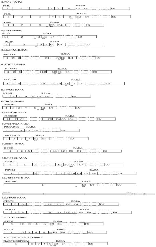
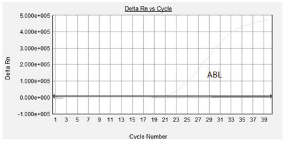
[0133] 1. The schematic diagram of the detection region and primer design region of the present invention is as follows figure 1 As shown, the reagents were prepared according to Table 1. The entire screening reagent was divided into 3 reaction tubes: the first and second reaction tubes were screening detection tubes for 14 fusion genes, and the third reaction tube was the internal reference gene ABL1 Detection tubes (APL-1+APL-2+ABL total three reaction tubes):

[0134] Table 1:

[0135]

[0136]

[0137]

[0138] 2. Adding samples: Add 2 μl of the cDNA in Example 2 to the PCR reaction solution of the detection system that has been packaged; directly add 2 μl of positive and negative control substances to the positive control and negative control; add 2 μl of normal saline to the blank control or not Any substance, the final reaction system is 25ul.

[0139] 3. Detection: The amplification program is as fol...


PUM


Abstract

The invention relates to a primer, a probe, a composition and a method for screening 14 fusion genes related to acute promyelocytic leukemia by using a real-time fluorescent quantitative PCR technology. The screened related fusion genes comprise PML-RAR[alpha], PLZF-RAR[alpha], NuMA-RAR[alpha], STAT5b-RAR[alpha], NPM-RAR[alpha], TBLR1-RAR[alpha], PRKAR1A-RAR[alpha], FNDC3B-RAR[alpha], BCOR-RAR[alpha], FIP1L1-RAR[alpha], IRF2BP2-RAR[alpha], STAT3-RAR[alpha], GRF2I-RAR[alpha] and NABP1-RAR[alpha]. The real-time fluorescence PCR technology is combined with the Tapman probe, common or uncommon APLdisease related fusion genes are integrated together for screening, diagnosis, treatment and prognosis monitoring of APL diseases can be assisted more comprehensively, and compared with previous methods such as FISH, the method has higher precision and facilitates result interpretation. Moreover, the primers and the probes are reasonably proportioned and optimized, and the detection conditions are optimized, so that the efficiency is greatly improved.

Description

technical field [0001] The invention belongs to the field of life science and biotechnology, and particularly relates to a method for designing specific primers and probes for fusion genes related to acute promyelocytes and using real-time fluorescent quantitative PCR technology for screening and the primers. APL-related fusion genes screened in the present invention include: PML-RARα, PLZF-RARα, NuMA-RARα, STAT5b-RARα, NPM-RARα, TBLR1-RARα, PRKAR1A-RARα, FNDC3B-RARα, BCOR-RARα, FIP1L1- RARα, IRF2BP2-RARα, STAT3-RARα, GTF2I-RARα, NABP1-RARα, a total of 14 fusion genes. This method is economical, convenient, and highly sensitive, and is suitable for screening and auxiliary diagnosis of clinical samples. [0002] technical background [0003] Acute promyelocytic leukemia (APL or M3) is a kind of acute leukemia with very dangerous clinical manifestations. The patients are usually young and middle-aged, and the incidence rate accounts for 10%-15% of acute non-lymphocytic leukemia...

Claims


Application Information

Patent Timeline
IPC IPC(8): C12Q1/6886C12Q1/6851C12N15/11
CPCC12Q1/6886C12Q1/6851C12Q2600/166C12Q2600/118
Inventor 杜翠邹媛董越张辰
Owner FUZHOU ADICON CLINICAL LAB INC
Why Eureka
Try Eureka