
Red fluorescent water-soluble cell nucleus targeting probe with V-shaped structure and application

A technology of red fluorescence and cell nucleus, which is applied in the field of targeted positioning of cell nucleus, amphiphilic/water-soluble fluorescent probe, and water-soluble cell nucleus targeting probe, which can solve the problems of unfavorable imaging and achieve good universality and imaging Simple and fast effect

Pending Publication Date: 2021-02-02
HUAZHONG UNIV OF SCI & TECH
View PDF11 Cites 5 Cited by
  • Summary
  • Abstract
  • Description
  • Claims
  • Application Information

AI Technical Summary

Problems solved by technology

[0003] The common dyes used for nuclear imaging are lipophilic dyes. Due to their inherent hydrophobicity, lipophilic dyes will spontaneously aggregate in physiological environments, and there will be strong background fluorescence during the imaging process, which is not conducive to imaging.

Method used


Image

  • Red fluorescent water-soluble cell nucleus targeting probe with V-shaped structure and application
  • Red fluorescent water-soluble cell nucleus targeting probe with V-shaped structure and application
  • Red fluorescent water-soluble cell nucleus targeting probe with V-shaped structure and application

Examples


Embodiment 1

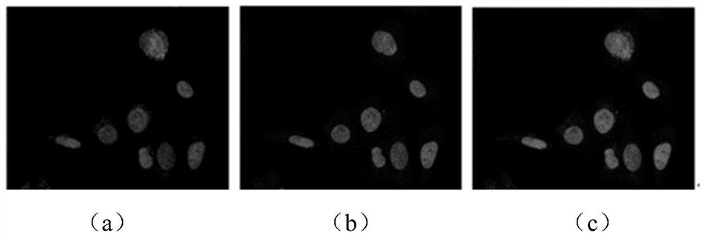
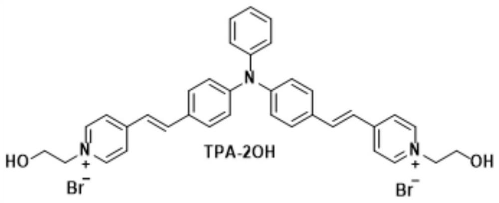
[0055] a structure such as figure 2 The fluorescent probe shown is abbreviated as TPA-2OH. Fluorescent imaging was performed by confocal microscopy by co-staining HeLa cells with TPA-2OH and the nuclear dye DAPI at a concentration of 2 μM for TPA-2OH and 5 μM for the nuclear dye DAPI.

[0056] by imaging figure 1 As shown in the red channel of TPA-2OH and the blue channel of the nuclear dye DAPI, obvious oval plaques can be seen. The imaging images of the two channels are superimposed, and the red part and the blue part overlap and become purple, which proves that TPA The -2OH nuclear dye DAPI targets the same cellular structure, the nucleus.

Embodiment 2

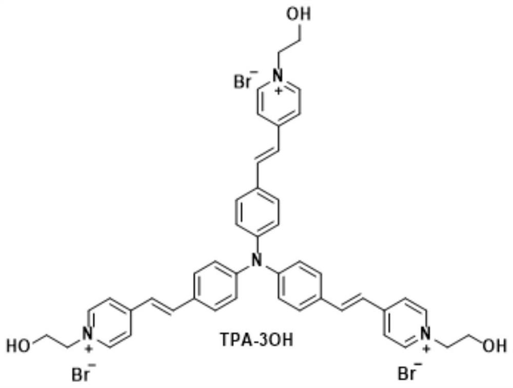
[0058] a structure such as image 3 The indicated fluorescent probes, whose names are abbreviated as TPA-3OH, were fluorescently detected by confocal microscopy by co-staining Hela cells with TPA-3OH and the nuclear dye DAPI at a concentration of 2 μM and nuclear dye DAPI imaging.

[0059] by imaging Figure 8 As shown, both the red channel of TPA-3OH and the blue channel of the nuclear dye DAPI can see obvious oval plaques. The imaging images of the two channels are superimposed, and the red part and the blue part overlap and become purple, which proves that TPA The -3OH nuclear dye DAPI targets the same cellular structure, the nucleus.

Embodiment 3

[0061] a structure such as Figure 4 For the indicated fluorescent probes, Hela cells were co-stained with the probe and the nuclear dye DAPI at a concentration of 2 μM of the probe molecule and 5 μM of the nuclear dye DAPI, and fluorescent imaging was performed by a confocal microscope.

[0062] by imaging Figure 9 As shown, both the red channel of the probe and the blue channel of the nuclear dye DAPI can see obvious oval plaques, and the imaging images of the two channels are superimposed, and the overlapping of the red part and the blue part becomes purple, which proves that the The probe molecule targets the same cellular structure as the nuclear dye DAPI, the nucleus.


PUM

PropertyMeasurementUnit
partition coefficientaaaaaaaaaa

Abstract

The invention belongs to the field of new materials, and discloses a red fluorescence water-soluble cell nucleus targeting probe with a V-shaped structure and application. The probe is an aggregation-induced emission fluorescence probe material with lipophilicity and hydrophilicity, the probe has a structural general formula shown as a formula (I) or a formula (II), M is a hydrophobic aromatic ring, and A is a hydrophilic nitrogen heterocyclic cation. According to the aggregation-induced emission fluorescent probe material, due to the particularity of the structure, the probe material has amphipathy and has better universality to the physiological environment of cells, the probe can independently target and position cell nucleuses and dye the cell nucleuses, background fluorescence is avoided, and no-clean imaging can be achieved.

Description

technical field [0001] The present invention belongs to the field of new materials, and more specifically relates to a red fluorescent water-soluble nucleus targeting probe with a V-shaped structure and its application. The probe is an amphiphilic / water-soluble fluorescent probe based on aggregation-induced luminescent properties Needles are especially useful for targeting cell nuclei in bioimaging. Background technique [0002] Cell fluorescence imaging has become an essential technology in modern cell biology. It can realize the fluorescence visualization of cells and their constituent structures, and has gradually become an effective tool for disease diagnosis and treatment. The nucleus is the largest and most important part of the cell. It is the cell metabolism, genetic information library and genetic control center. The genetic material deoxyribonucleic acid and ribonucleic acid are mainly stored in the nucleus. DNA damage has been linked to a range of diseases, inclu...

Claims


Application Information

Patent Timeline
Patent Type & Authority Applications(China)
IPC IPC(8): C07D213/38C07D401/14C07D409/14C07D417/14C09K11/06C09B57/00G01N21/64
CPCC07D213/38C07D401/14C07D409/14C07D417/14C09K11/06C09B57/00C09B23/145C09B23/141C09B21/00G01N21/6428C09K2211/1014C09K2211/1029C09K2211/1037C09K2211/1092G01N2021/6439
Inventor 朱明强徐楚燃李冲王亚龙
Owner HUAZHONG UNIV OF SCI & TECH
Learn More