Construction method and application of glioma cell line suitable for two-photon living imaging

A glioma cell and in vivo imaging technology, which is applied in the construction and application of two-photon in vivo imaging glioma cell lines, to achieve the effect of improving accuracy

Pending Publication Date: 2021-02-02
LANZHOU UNIVERSITY
View PDF3 Cites 1 Cited by
  • Summary
  • Abstract
  • Description
  • Claims
  • Application Information

AI Technical Summary

Problems solved by technology

However, this technology has certain requirements for the sample to be imaged, that i

Method used


Image

  • Construction method and application of glioma cell line suitable for two-photon living imaging
  • Construction method and application of glioma cell line suitable for two-photon living imaging
  • Construction method and application of glioma cell line suitable for two-photon living imaging

Examples


Embodiment 1

[0036] Construction of a glioma cell line (GL261-ZsGreen) expressing green fluorescence:

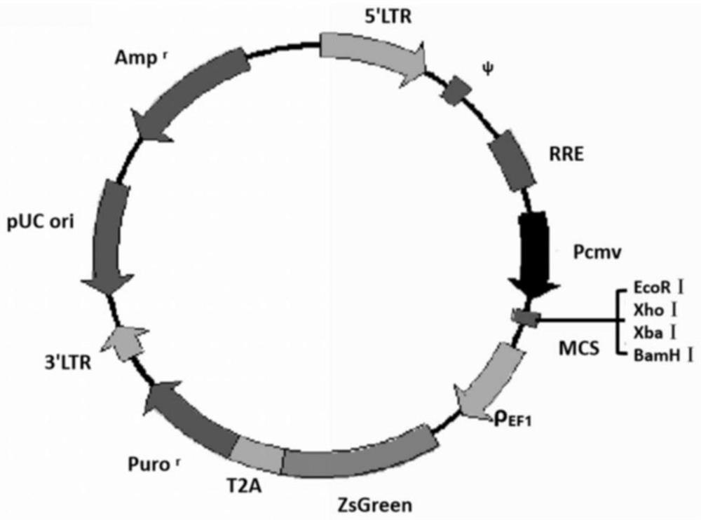
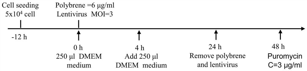
[0037] Step 1. Spread 5×10 cells in a 24-well culture plate 12 hours in advance 4 GL261 glioma cells, after the cell confluency reaches about 60%, add 6 μg / ml polybrene transfer agent to the DMEM medium, and then use ZsGreen fluorescence with CMV promoter and two-photon excitation band The pHBLV-CMV-MCS-EF1-ZsGreen-T2A-puro lentivirus with reporter gene and puromycin resistance gene was used to infect the pre-laid GL261 glioma cells;

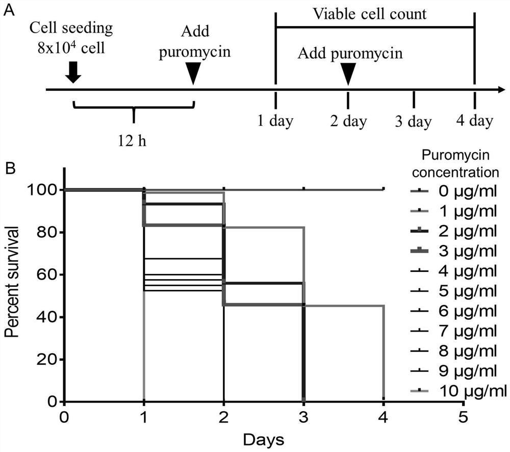
[0038] Step 2. After the above-mentioned GL261 glioma cells infected with the lentivirus were grown for 48 hours, replace with fresh DMEM medium containing puromycin at a concentration of 2 μg / ml, and screen the GL261 glioma cells. Replace with fresh medium containing puromycin once every day;

[0039] Step 3. The glioma cells with green fluorescent label (GL261-ZsGreen) screened in step 2 are monoclonally amplified and stored frozen.

[0040] Construct...

Embodiment 2

[0046] Construction of a glioma cell line (GL261-ZsGreen) expressing green fluorescence:

[0047] Step 1. Spread 5×10 cells in a 24-well culture plate 12 hours in advance 4 A GL261 glioma cell, after the cell confluency reaches about 60%, add 7 μg / ml polybrene transfer agent to the DMEM medium, and then use ZsGreen fluorescence with CMV promoter and two-photon excitation band The pHBLV-CMV-MCS-EF1-ZsGreen-T2A-puro lentivirus with reporter gene and puromycin resistance gene was used to infect the pre-laid GL261 glioma cells;

[0048] Step 2. After the above-mentioned GL261 glioma cells infected with lentivirus were grown for 48 hours, fresh DMEM medium containing puromycin at a concentration of 2 μg / ml was replaced to screen GL261 glioma cells. Replace with fresh medium containing puromycin once every day;

[0049] Step 3. The glioma cells with green fluorescent label (GL261-ZsGreen) screened in step 2 are monoclonally amplified and stored frozen.

Embodiment 3

[0051] Construction of a glioma cell line (GL261-ZsGreen) expressing green fluorescence:

[0052] Step 1. Spread 5×10 cells in a 24-well culture plate 12 hours in advance 4 GL261 glioma cells, after the cell confluency reaches about 60%, add 8 μg / ml polybrene transfer agent to the DMEM medium, and then use ZsGreen fluorescence with CMV promoter and two-photon excitation band The pHBLV-CMV-MCS-EF1-ZsGreen-T2A-puro lentivirus with reporter gene and puromycin resistance gene was used to infect the pre-laid GL261 glioma cells;

[0053] Step 2. After the above-mentioned GL261 glioma cells infected with lentivirus were grown for 48 hours, fresh DMEM medium containing puromycin concentration of 3 μg / ml was replaced to screen GL261 glioma cells. Replace with fresh medium containing puromycin once every day;

[0054] Step 3. The glioma cells with green fluorescent label (GL261-ZsGreen) screened in step 2 are monoclonally amplified and stored frozen.


PUM

PropertyMeasurementUnit
Thicknessaaaaaaaaaa

Abstract

The invention discloses a construction method of a glioma cell line suitable for two-photon living imaging. The construction method comprises the following steps: constructing a glioma cell in-situ transplantation animal model through glioma cells, and researching the dynamic development change of the glioma cells in the brain of a mouse by virtue of a two-photon living body imaging technology. The GL261-ZsGreen glioma cells obtained through transformation can be successfully used for constructing a glioma in-situ transplantation living animal model, the positions of tumor cells can be clearlyobserved in the imaging observation process, cell-level examination can be accurately performed when the GL261-ZsGreen glioma cells are applied to the research field, the glioma boundary can be obviously distinguished, and a potential means is provided for effectively and accurately excising tumors. Each proliferated GL261-ZsGreen cancer cell is provided with a green fluorescence label, so that single cancer cells subjected to dispersive metastasis can be accurately positioned, and a possible scheme is provided for further researching the diffusion of cancer cells and the accurate treatment of glioma.

Description

technical field [0001] The invention belongs to the technical fields of biology and oncology, and in particular relates to the construction and application of a glioma cell line suitable for two-photon living imaging. Background technique [0002] Glioma is the most common primary tumor in the central nervous system. It has the characteristics of high recurrence, poor prognosis, and high mortality of patients, which seriously threatens the survival time and quality of patients. There is no particularly effective treatment strategy. In recent years, the incidence of brain tumors has been on the rise. Many brain tumors, especially astrocytomas, are difficult to treat thoroughly through surgery after they are diagnosed, and the prognosis is also very poor. Therefore, glial Early diagnosis and treatment of glioma play a vital role in improving the survival of glioma patients. [0003] The current clinical application of glioma detection and diagnosis technology mainly relies o...

Claims


Application Information

Patent Timeline
IPC IPC(8): C12N5/10C12N15/867C12N15/65A01K67/027A61K49/00
CPCC12N5/0693C12N15/86C12N15/65A01K67/0271A61K49/0047C12N2740/15043A01K2227/105A01K2267/0331
Inventor 张胜祥谢文广杨婧张娜
Owner LANZHOU UNIVERSITY
Why Eureka
Try Eureka