Methods for growing african swine fever virus in fetal porcine lung alveolar macrophage cells
A technology of alveolar macrophages and macrophages, applied in the field of growing African swine fever virus in fetal pig alveolar macrophages, can solve the problem of reducing the quantity and quality of pork, lack of ASF disease diagnosis technology, and high mortality and morbidity issues
- Summary
- Abstract
- Description
- Claims
- Application Information
AI Technical Summary
Problems solved by technology
Method used
Image
Examples
Embodiment 1
[0188] Example 1: Cell material and cell growth
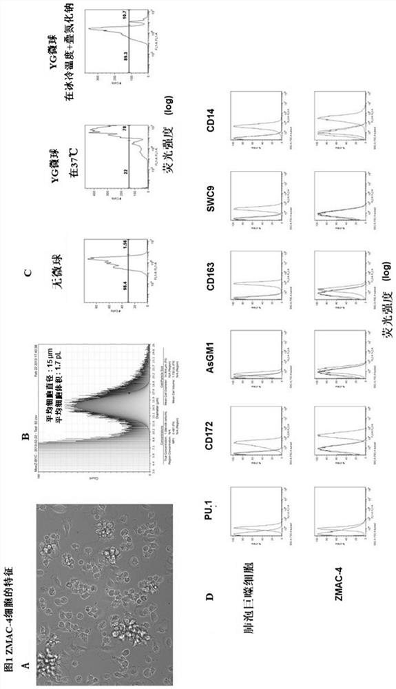
[0189] cell material : Cells designated ZMAC-1 were prepared from a porcine alveolar macrophage cell line and deposited under the Budapest Treaty with the American Type Culture Collection (ATCC; 10801 University Avenue, Manassas, Virginia), a recognized international depository institution state, United States). Cells characterized as derived from Sus scrofa (pig / pig) lung tissue have accession number ATCC patent deposit number PTA-8764. Pursuant to the ATCC Deposit Document dated December 7, 2007 (Budapest Treaty on the International Recognition of the Deposit of Microorganisms for the Purposes of Patent Procedure; International Forms; Issue of Receipt of Original Deposit under Article 7.3 and Issue of Viability Statement under Article 10.2), The date of culture receipt for patent deposit designation PTA-8764 is November 14, 2007.
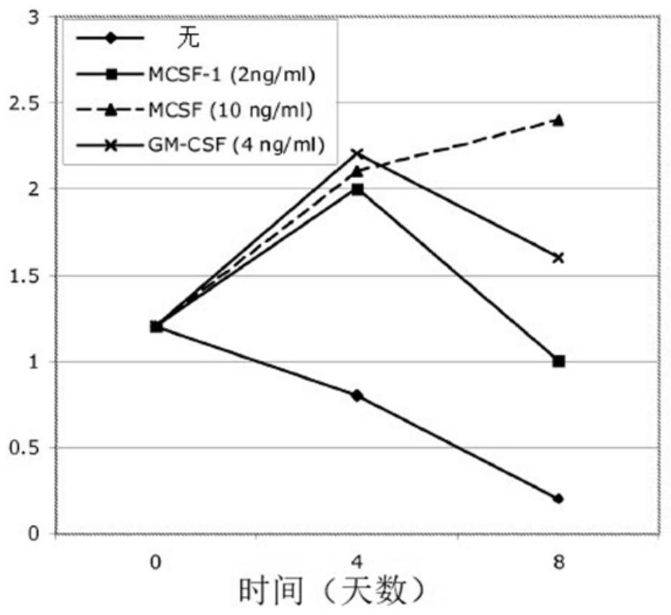
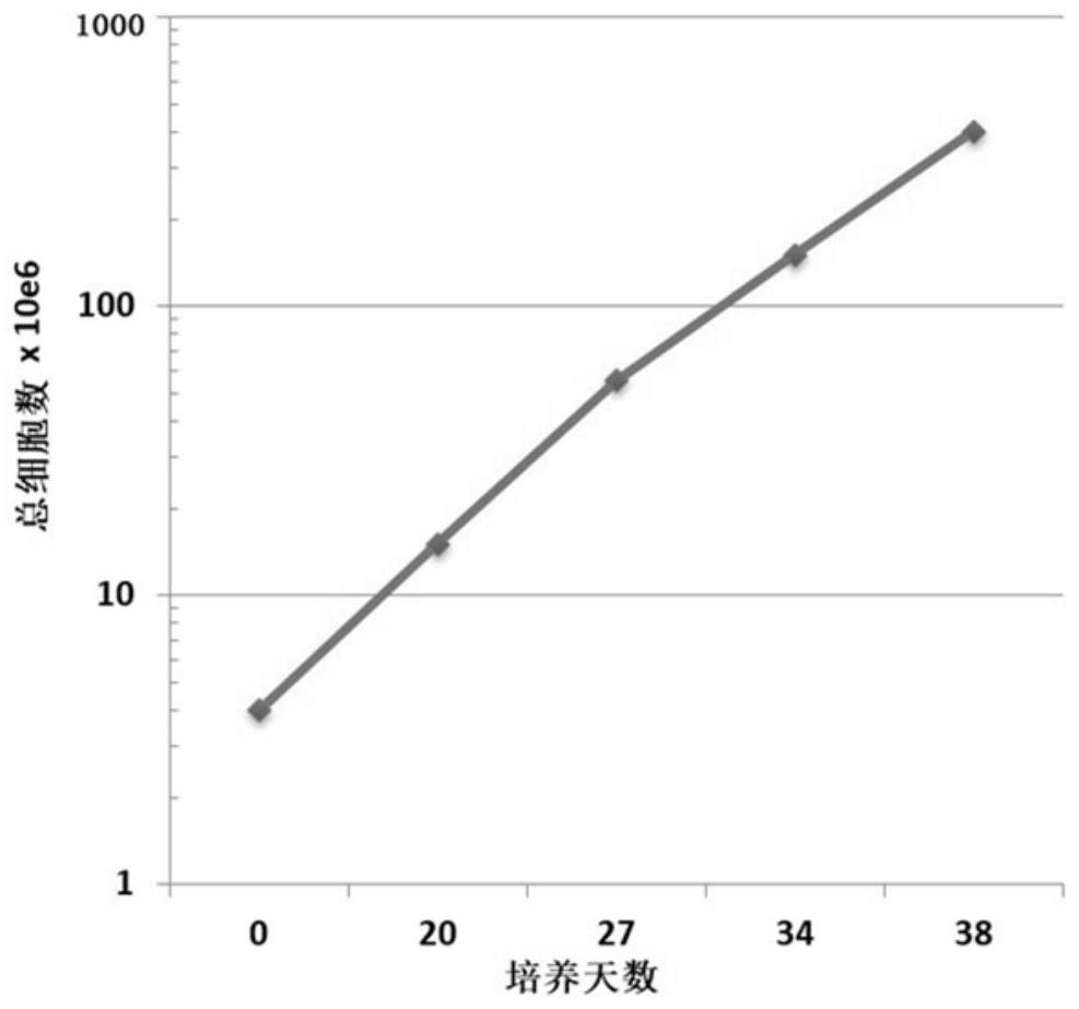
[0190] cell growth : In some embodiments, cells such as ZMAC-1 cells are cultured in vit...
Embodiment 2
[0193] Example 2: Production of cellular material and method of isolation from fetal pig samples
[0194] Further compositions and methods were developed. In an independent attempt, material was obtained from a 60-day gestation sow (identified as sow number 5850) in the swine herd at the University of Illinois Veterinary Research Farm. After euthanasia, the uteri were aseptically removed from the abdominal cavity and transferred to a cell culture laboratory. Usually performed under sterile conditions using a biosafety console. Six fetuses were aseptically harvested from the uterus and placed in plastic Petri dishes. The lung organ with intact trachea connecting to the lung is dissected from the heart, esophagus and other membranes. The exterior of the lungs was rinsed thoroughly with sterile Hank's Balanced Salt Solution (HBSS) to remove any visible blood and other contaminated residual tissue. Bronchoalveolar lavage was performed by placing the lungs in a clean sterile Pe...
Embodiment 3
[0199] Example 3: Establishment of ZMAC cell cultures and testing for ASFV susceptibility at the Pirbright Institute
[0200] Vials of ZMAC cells provided by Aptimmune Biologics, Inc. (1005 N. Warson Road, Suite 305, St. Louis, MO 63132) were revived from liquid nitrogen storage. Briefly, cells were quickly thawed at 37°C (i.e., degrees Celsius), added to 10 ml of warm medium, centrifuged at 330xg and 4°C for 7 min to remove freezing medium, and then incubated at T75 ultra-low attachment ( ULA) culture flasks were cultured in a total of 50 ml of medium and 5 ng / ml MCSF. After 3 days, the cultures were re-fed with new medium at a 1 :2 dilution with supplemented M-CSF at a final concentration of 5 ng / ml in the total volume (100 ml). Additional refeeds were performed every three to four days with fresh medium (prepared less than 7 days earlier) at a dilution of 1 :2 culture volume with a final M-CSF supplementation of 5 ng / ml. When the culture reached approximately 150 ml in th...
PUM
 Abstract
Description
Claims
Application Information
 - R&D
- Intellectual Property
- Life Sciences
- Materials
- Tech Scout
- Unparalleled Data Quality
- Higher Quality Content
- 60% Fewer Hallucinations
Browse by: Latest US Patents, China's latest patents, Technical Efficacy Thesaurus, Application Domain, Technology Topic, Popular Technical Reports.
© 2025 PatSnap. All rights reserved.Legal|Privacy policy|Modern Slavery Act Transparency Statement|Sitemap|About US| Contact US: help@patsnap.com


