A paper-based detection method for exosomes

A detection method, exosome technology, applied in the direction of biochemical equipment and methods, microbial measurement/inspection, etc., to achieve the effect of low requirements, enhanced visual signal, and easy detection and operation

Active Publication Date: 2021-06-11
北京红果源生物技术有限公司
View PDF0 Cites 0 Cited by
  • Summary
  • Abstract
  • Description
  • Claims
  • Application Information

AI Technical Summary

Problems solved by technology

Compared with antibodies, nucleic acid aptamers have the advantages of stable chemical properties, easy preparation, easy modification, and easy combination with nucleic acid amplification technology for signal amplification. However, there is no report on the use of nucleic acid LFB for exosome detection.

Method used


Image

  • A paper-based detection method for exosomes
  • A paper-based detection method for exosomes
  • A paper-based detection method for exosomes

Examples


Embodiment 1

[0063] Example 1 Establishment of a paper-based detection method for exosomes

[0064] 1. Experimental materials

[0065] 20×SSC, Tris-HCl, sodium phosphate dodecahydrate, bovine serum albumin (BSA), Tween-20, sucrose and TMB chromogenic solution, etc.

[0066] The sequence design is as follows (SEQ ID NO:1-4):

[0067]

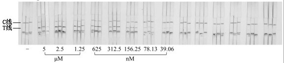
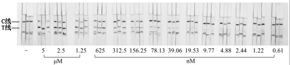
[0068] 2. LFB detection and verification of single-stranded DNA targets

[0069] Dilute the single-stranded DNA target 2-fold concentration gradient, add 50 µL system buffer and 1 µL single-stranded DNA target solution to the sample pool, and drop-coat 1 µL PtAu-NiO on the superposition of the test paper sample pad and the binding pad 2 After the nanozyme signal probe, insert the test paper sample pad into the sample pool, wait for 3 minutes, and observe the result with the naked eye. When both the C line and the T line are black, it is determined to be positive. The color of the T line becomes lighter as the concentration of the ssDNA target decreases,...

Embodiment 2

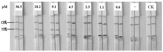
[0078] Example 2 Practical investigation of detection method

[0079] According to the detection method constructed in Example 1, exosomes extracted from equal volumes of HepG2 and L02 cell supernatants were detected. LFB can effectively distinguish tumor exosomes from non-tumor exosomes ( Figure 6 ).

Embodiment 3

[0080] Embodiment 3 Amplification effect verification of detection method

[0081] After conversion and amplification of exosomes, the gray pixel area of ​​each T line in the LFB detection result was substituted into the gray pixel area of ​​the T line obtained by LFB detection after 2-fold gradient dilution of the single-stranded DNA target, and the standard curve fitting was obtained. Formula: y =14.829e 0.0005x In , the concentration of the single-stranded DNA target after exosome conversion and amplification was obtained. The exosome concentration was converted to the concentration of the substance and compared with the single-stranded DNA target. The molecular magnification of exosomes by this method is 1000 times (Table 1).

[0082] Table 1 Molecular magnification of exosomes after target conversion and amplification

[0083]


PUM

PropertyMeasurementUnit
Sensitivityaaaaaaaaaa

Abstract

The present invention provides a paper-based detection method for exosomes, including: (1) recognition and molecular conversion system, (2) amplification reaction system, and (3) LFB detection and analysis system. The present invention converts exosomes into single-stranded DNA targets, amplifies the single-stranded DNA through nucleic acid amplification technology, and further utilizes PtAu-NiO 2 The amplified single-stranded DNA is detected by the lateral flow chromatography test paper with the nanozyme as the signal probe. The invention uses nucleic acid lateral flow chromatography test paper as the terminal detection platform, solves the problem that exosomes cannot be detected qualitatively and quantitatively at the same time, and realizes simple and low-cost detection of exosomes.

Description

technical field [0001] The invention belongs to the technical field of biological detection, and in particular relates to a paper-based detection method for exosomes. Background technique [0002] The nanoscale particle size and ultra-trace concentration of exosomes in body fluids pose great challenges for their identification and analysis. Existing analysis methods not only rely on large and expensive instruments, but also require complex pretreatment processes and professional operators. In addition, traditional exosome detection methods cannot be qualitative and quantitative at the same time. Therefore, the development of fast, simple, low-cost, and highly sensitive exosome detection methods is the key to the rapid development and popularization of clinical disease diagnosis and treatment methods using exosomes as biomarkers. [0003] Lateral Flow Chromatography (LFB) is a sensitive, cheap and simple visual rapid detection method. Early LFBs were based on immune recogni...

Claims


Application Information

Patent Timeline
Patent Type & Authority Patents(China)
IPC IPC(8): C12Q1/6851
CPCC12Q1/6851C12Q2525/205C12Q2563/155C12Q2565/625C12Q2565/519C12Q2545/114C12Q2563/137
Inventor 许文涛黄昆仑王馨娴李舒婷程楠朱龙佼杜再慧张博洋
Owner 北京红果源生物技术有限公司
Why Eureka
Try Eureka