Diagnosis method for bone metastasis tumor in nuclide bone imaging based on deep learning

A technology of deep learning and diagnostic methods, applied in the field of medical image processing, can solve problems such as high complexity of image acquisition, easy misdiagnosis, and difficulties for doctors

Pending Publication Date: 2021-04-09
SHANGHAI TENTH PEOPLES HOSPITAL
View PDF6 Cites 4 Cited by
  • Summary
  • Abstract
  • Description
  • Claims
  • Application Information

AI Technical Summary

Problems solved by technology

[0004] At present, the analysis of whole-body bone scan images mainly relies on the manual reading of nuclear medicine physicians, and according to the physician's personal experience, the diagnosis is made by checking the whole-body bone scan images to find abnormal lesion locations. Physicians have to do a lot of repetitive work for this
Due to the high complexity of image acquisition, the great variability among patients, the poor image quality, and the large subjective factors in manual film reading, there are deviations in the analysis and diagnosis results of whole-body bone scan images, which are prone to misdiagnosis

Method used


Image

  • Diagnosis method for bone metastasis tumor in nuclide bone imaging based on deep learning
  • Diagnosis method for bone metastasis tumor in nuclide bone imaging based on deep learning
  • Diagnosis method for bone metastasis tumor in nuclide bone imaging based on deep learning

Examples


Embodiment Construction

[0038] The present invention will be further described below in conjunction with specific examples, so as to better understand the present invention.

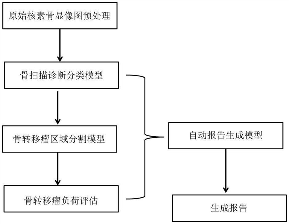
[0039] 1. Data processing

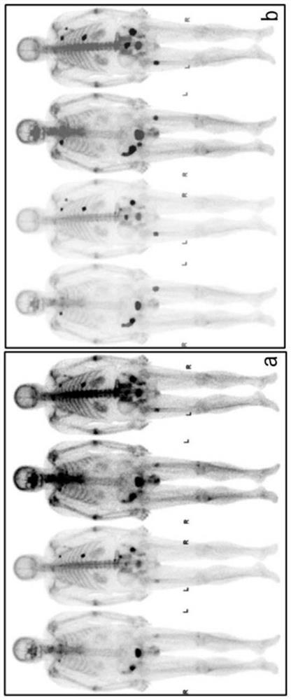
[0040] The original data contains the front and back images under two different gray values, that is, one piece of data corresponds to four sub-images, such as figure 2 as shown in a. Two professional nuclear medicine physicians collaborated to diagnose bone metastases and use Labelme to outline the bone metastases and the bladder area in the image. The labeling results are as follows figure 2 as shown in b. The original image size is unified to 1024×1024 by means of bilinear difference. Randomly divide 80% as the training set and the remaining 20% ​​as the test set.

[0041] 2. Bone scan diagnostic classification model

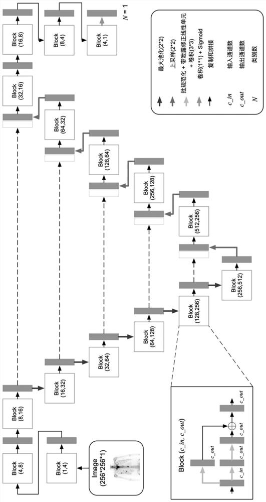
[0042] 2.1 Data processing

[0043] The diagnostic classification model uses a 256×256 image as input, that is, a 1024×1024 image can get 28 subimages of 256×256. Segmentatio...


PUM


Abstract

The invention provides a method for diagnosing bone metastases in nuclide bone imaging based on deep learning. The method relates to a bone scanning diagnosis classification model, a bone metastasis tumor region segmentation model and a bone metastasis tumor load evaluation and automatic report generation model. By the adoption of the method, the bone metastasis tumor can be judged, automatic region segmentation can be conducted, the recognition accuracy is high, and full-automatic analysis from original image input to report generation is preliminarily achieved.

Description

technical field [0001] The invention relates to the technical field of medical image processing, in particular to a method for diagnosing bone metastases in nuclide bone imaging based on deep learning. Background technique [0002] The incidence of bone metastases is 35 to 40 times that of primary bone tumors. Cancer bone metastasis is one of the main causes of cancer pain. It causes pathological fractures, spinal cord compression, hypercalcemia and bone marrow failure and other complications, which accelerate the development of the disease and seriously affect the quality of life of cancer patients. In the past ten years, many disciplines have made unremitting efforts in the pathogenesis, prevention and treatment of bone metastases, but so far no effective cure has been found. [0003] At present, bone scan is the most commonly used examination method for the analysis of bone metastases and the evaluation of curative effect. The radionuclide used 99m Tc-MDP is imaged by t...

Claims


Application Information

Patent Timeline
IPC IPC(8): G16H50/50G16H50/30G16H15/00G06T7/00G06T7/11G06K9/62G06N3/04G06N3/08
CPCG16H50/50G16H50/30G16H15/00G06T7/0012G06T7/11G06N3/08G06T2207/20081G06T2207/20084G06T2207/30008G06T2207/30096G06N3/045G06F18/214G06F18/24
Inventor 李丹刘思敏冯明吕中伟王胤
Owner SHANGHAI TENTH PEOPLES HOSPITAL
Why Eureka
Try Eureka