Malignant melanoma biomarker PTGER2 and application thereof
A melanoma and drug technology, applied in the field of biomedicine, can solve problems such as the current situation is not optimistic, and there is no significant progress in clinical treatment
- Summary
- Abstract
- Description
- Claims
- Application Information
AI Technical Summary
Problems solved by technology
Method used
Image
Examples
Embodiment 1
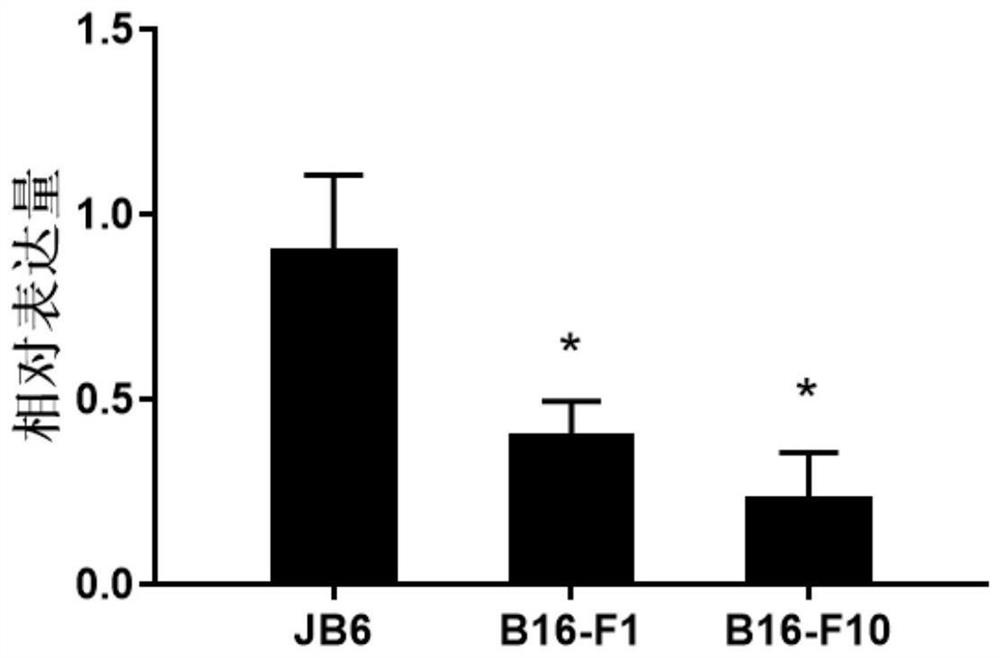
[0027] The mRNA levels of PTGER2 in mouse normal epidermal cell JB6, mouse melanoma high-metastatic cell B16-F10 and mouse melanoma low-metastatic cell B16-F1 were compared.
[0028] Mouse melanoma cells B16-F10 were inoculated in DMEM medium containing 10% fetal bovine serum (adding penicillin and streptomycin 100 U / mL), placed at 37°C, 5% CO 2 Cultured in an incubator, digested and passaged with 0.25% trypsin.
[0029] The operation steps are as follows:
[0030] RNA extraction and fluorescent quantitative PCR detection: Use the total RNA rapid extraction kit (Flying Biotech) to extract the RNA of the cells, and perform reverse transcription according to the conventional steps to form cDNA, and then detect the expression effect of PTGER2 by real-time fluorescent quantitative PCR. Among them, The sequences of cDNA amplification primer pairs are:
[0031] qPCR primer F: 5'-CAGCTCGGTGATGTTCTCGG-3' (SEQ ID NO: 1);
[0032] qPCR primer R: 5'-GAGCACCAATTCCGTTACCAG-3' (SEQ ID NO...
Embodiment 2
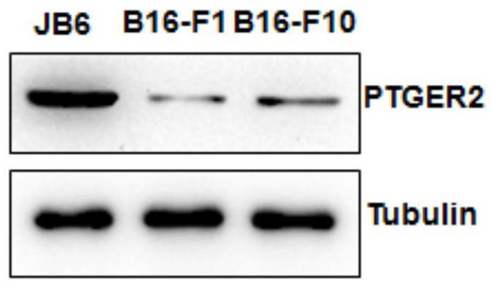
[0035] The protein levels of PTGER2 in mouse normal epidermal cells and mouse melanoma high-metastatic cells B16-F10 and mouse melanoma low-metastatic cells B16-F1 were compared.
[0036] The operation steps are as follows:
[0037] Collect the cells at 80% confluence, discard the supernatant after centrifugation, rinse twice with PBS, and discard the supernatant. Add RIPA lysate and lyse on ice for 20 min. The supernatant was collected by centrifugation at 12000 g for 10 min. Add 1xSDS sample buffer, mix well by pipetting and then boil for denaturation for 5min. Total proteins were separated by 10% SDS-PAGE gel and then transferred to PVDF membrane. 5% BSA was blocked at room temperature for 2 hours, incubated with PTGER2 antibody (GeneTex, GTX66706 (1:1000 dilution)) overnight at 4°C, and washed 3 times with TBST. The secondary antibody was incubated at room temperature for 1 h, and washed 3 times with TBST. ECL ultra-sensitive chemiluminescent solution was developed an...
Embodiment 3
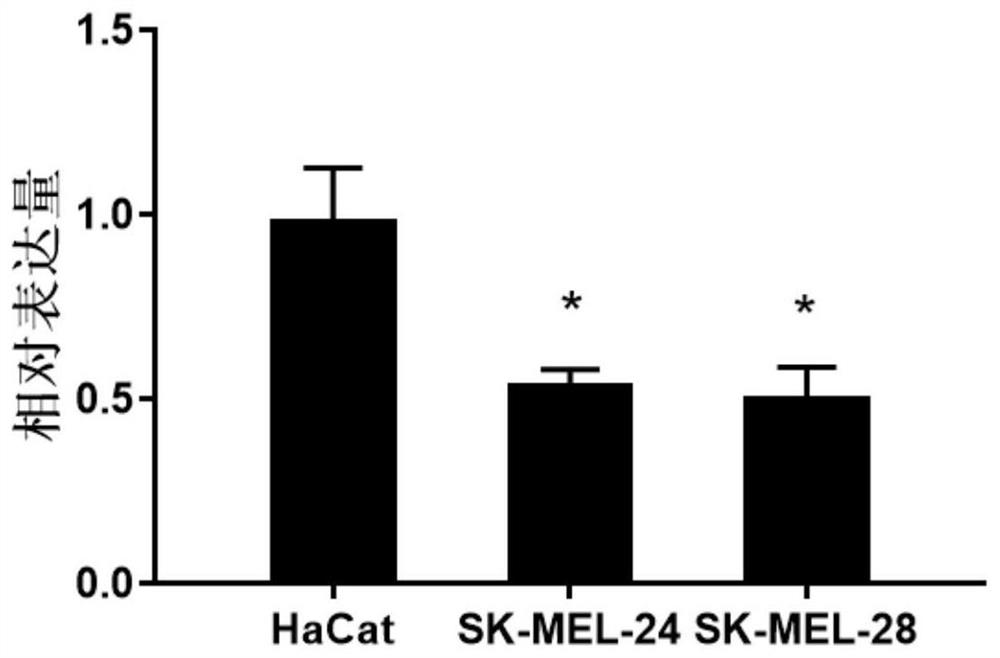
[0040] The expression levels of PTGER2 in human normal epidermal cells HaCat and human melanoma cells SK-MEL-24 and SK-MEL-28 were compared.
[0041] For specific steps, refer to the experimental method described in Example 1.
[0042] The sequences of cDNA amplification primer pairs are:
[0043] qPCR primer F: 5'-CGATGCTCATGCTCTTCGC-3' (SEQ ID NO: 3);
[0044] qPCR primer R: 5'-GGGAGACTGCATAGATGACAGG-3' (SEQ ID NO: 4).
[0045] The result is as image 3 As shown, the expression level of PTGER2 was found to be lower in human melanoma cells than in normal epidermal cells.
PUM
 Abstract
Description
Claims
Application Information
 - R&D
- Intellectual Property
- Life Sciences
- Materials
- Tech Scout
- Unparalleled Data Quality
- Higher Quality Content
- 60% Fewer Hallucinations
Browse by: Latest US Patents, China's latest patents, Technical Efficacy Thesaurus, Application Domain, Technology Topic, Popular Technical Reports.
© 2025 PatSnap. All rights reserved.Legal|Privacy policy|Modern Slavery Act Transparency Statement|Sitemap|About US| Contact US: help@patsnap.com


