A recombinant fusion protein smsap of Schistosoma mansoni and its application in immunodiagnosis of schistosomiasis
A technology of Schistosoma mansoni and fusion protein, which is applied in the biological field, can solve problems such as differences, and achieve the effects of good repeatability, good specificity, and high sensitivity of results
- Summary
- Abstract
- Description
- Claims
- Application Information
AI Technical Summary
Problems solved by technology
Method used
Image

Examples
Embodiment 1
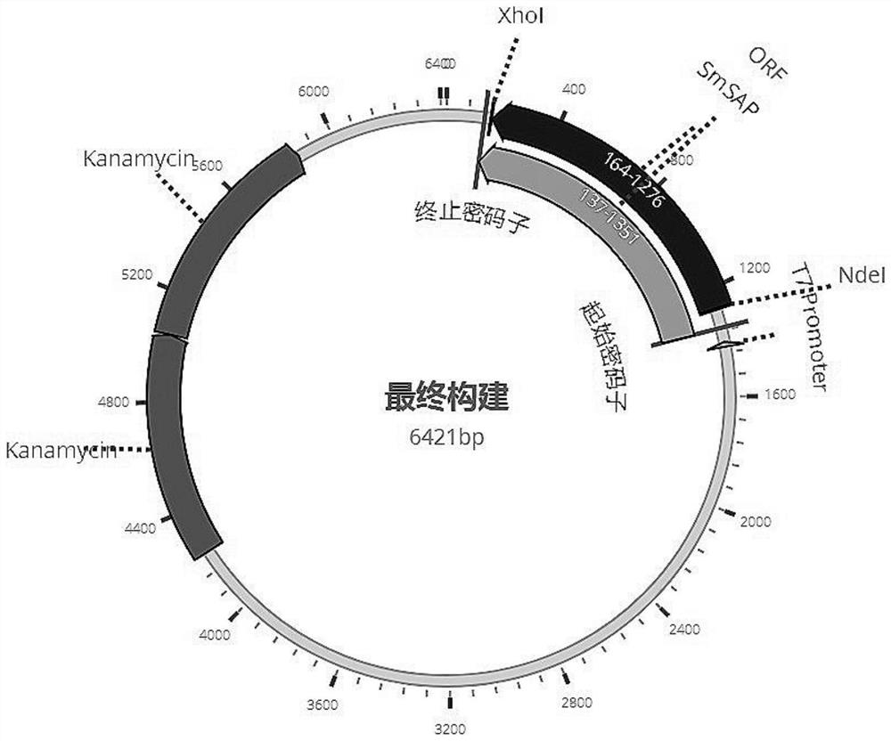
[0027] Example 1 Schistosoma mansoni SmSAP fusion protein gene synthesis and prokaryotic expression vector construction
[0028] The SmSAP gene encoded by the whole genome of Schistosoma mansoni is predicted by bioinformatics methods. The amino acid sequence of the SmSAP fusion protein of Schistosoma mansoni is shown in the sequence table SEQ ID NO.1. The SmSAP fusion protein is composed of Schistosoma mansoni with the signal peptide sequence removed Saposin-like protein 1 (SmSAP1) and Schistosoma mansoni saposin-like protein 2 (SmSAP2), the amino acid sequences of SmSAP1 protein and SmSAP2 protein are as shown in the sequence table SEQ ID NO.2 and SEQ ID NO.3 . According to the codon preference of Escherichia coli, the SmSAP fusion protein coding gene was designed, and then the SmSAP fusion gene of Schistosoma mansoni was prepared by gene synthesis technology, and its nucleotide sequence was shown in SEQ ID NO.4 in the sequence table. Nde I and Xho I restriction sites were d...
Embodiment 2
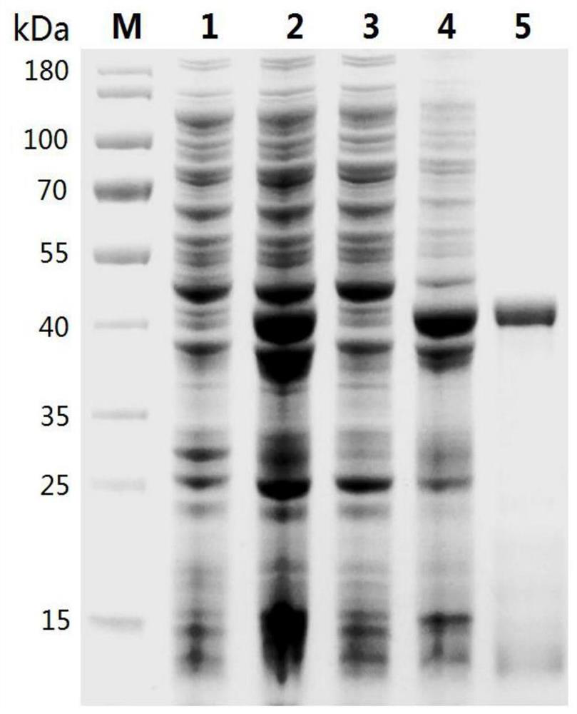
[0030] Example 2 Prokaryotic expression and purification of Schistosoma mansoni SmSAP fusion protein
[0031] Transform the pET28a-SmSAP recombinant plasmid with correct sequencing into expression competent cells Transetta (DE3), spread the transformed competent cells on LB medium plates (containing 50 μg / ml kanamycin), and culture overnight at 37°C; Positive clones were inoculated into 15 mL of LB liquid medium (containing 50 μg / ml kanamycin), cultured overnight at 37 ° C, and transferred 10 mL of medium into 1L LB medium (containing 50 μg / ml kanamycin) the next day, Continue to culture to OD 600nm When the value is 0.8, IPTG with a final concentration of 1 mM was added to induce expression for 4 hours, the cells were collected by centrifugation, and frozen at -80°C for future use.
[0032] Take a small amount of pre-induction and post-induction bacteria resuspended in PBS buffer, add SDS-PAGE sample buffer, mix well, boil in boiling water bath for 5min to denature the prote...
Embodiment 3
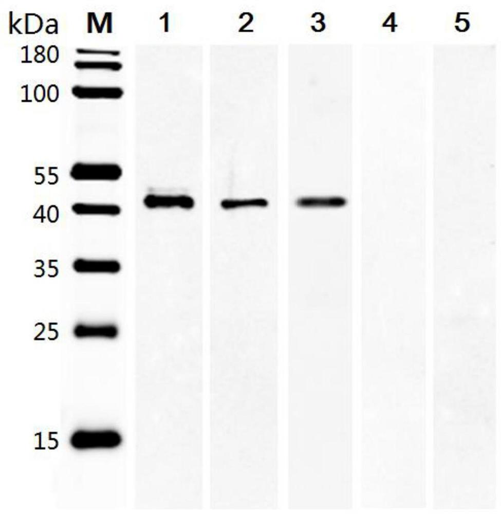
[0035] Example 3 Antigenicity Detection of Schistosoma mansoni SmSAP Recombinant Protein
[0036] Take 200ng of SmSAP recombinant protein as a sample, and perform SDS-PAGE gel electrophoresis; transfer the protein in the PAGE gel to PVDF membrane by wet transfer method; seal the PVDF membrane with 5% skim milk powder at room temperature for 2 hours, and wash 3 times with TBST buffer; Using mouse anti-His tag antibody as positive control and healthy human serum as negative control, add serum from patients with Schistosomiasis mansoni, Schistosomiasis haematobium and Schistosomiasis japonicum patients (diluted with blocking solution 1:100), 4°C Incubate overnight, wash 3 times with TBST buffer; add fluorescently labeled anti-mouse IgG antibody or anti-human IgG antibody (diluted with blocking solution 1:10,000), incubate at 37°C for 1 hour in the dark, wash 3 times with TBST buffer; use Odyssey infrared laser imaging system scanning imaging. The result is as image 3 As shown,...
PUM
| Property | Measurement | Unit |
|---|---|---|
| Sensitivity | aaaaa | aaaaa |
Abstract
Description
Claims
Application Information
 - R&D
- Intellectual Property
- Life Sciences
- Materials
- Tech Scout
- Unparalleled Data Quality
- Higher Quality Content
- 60% Fewer Hallucinations
Browse by: Latest US Patents, China's latest patents, Technical Efficacy Thesaurus, Application Domain, Technology Topic, Popular Technical Reports.
© 2025 PatSnap. All rights reserved.Legal|Privacy policy|Modern Slavery Act Transparency Statement|Sitemap|About US| Contact US: help@patsnap.com