

Application of a Nanoscale Coordination Polymer in Nuclear Magnetic Imaging
A coordination polymer and nuclear magnetic resonance imaging technology, which is applied in the field of imaging diagnostic contrast agents, can solve the problems that the sensitivity of identification is difficult to meet clinical needs, increase contrast and imaging sensitivity, and shorten the relaxation time of hydrogen protons in water, etc.
- Summary
- Abstract
- Description
- Claims
- Application Information
AI Technical Summary
Problems solved by technology
Method used
Image

Examples
Embodiment 1
[0034] Example 1 Preparation of Nanoscale Coordination Polymer as Nuclear Magnetic Imaging Enhancer
[0035] Prepare GdCl first 3 ·6H 2 100 mL of O (10.0 mM, pH=7.4) solution, 100 mL of 5'-GMP (10.0 mM, pH=7.4) solution and 100 mL of Hemin (1.0 mM, pH=7.4) HEPES buffer were used for later use. Take 20mL 5'-GMP (10.0mM, pH = 7.4) in a 100mL beaker, add 20mL Hemin (1.0mM, pH = 7.4) HEPES buffer, stir magnetically for 10 minutes, then add 30mL GdCl to the system 3 ·6H 2 O (10.0 mM, pH=7.4), continue magnetic stirring for 30 minutes to form dark brown nanoparticle aggregates. Separation by centrifugation (12000rpm × 10 min) to obtain nanoscale crude product, and then wash with deionized water (10mL × 3 times) to remove free molecular monomers, and separation by centrifugation (12000rpm × 10min) to obtain nanoscale pure solid product, and finally add 10mL of normal saline was ultrasonically resuspended to obtain the nanoscale radiosensitizing drug Hemin@Gd-NCPs, the concentrati...
Embodiment 2
[0036] Example 2 Characterization of Longitudinal Relaxation Rate of Nanoscale Coordination Polymer Hemin@Gd-NCPs as NMR Enhancer and Enhancement Effect of NMR Imaging in Vivo
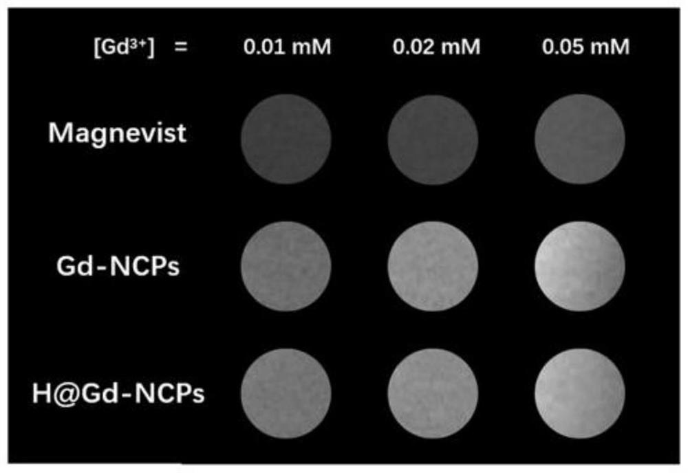
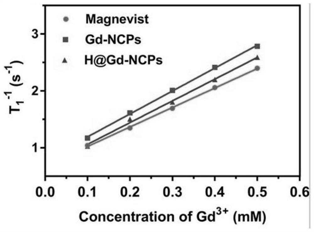
[0037] Configure Gd 3+ Magnevist, Gd-NCPs, and Hemin@Gd-NCP samples with a concentration of 0.01mM, 0.02mM, and 0.05mM were photographed using a nuclear magnetic imaging instrument (Biospec 7T / 20USR, Germany) at a magnetic field strength of 7.0Tesla(T) T1-weighted imaging and quantification of individual samples. In order to evaluate the accumulation of Hemin@Gd-NCPs in the tumor body and the ability of nuclear magnetic imaging and compare it with the clinically applied nuclear magnetic imaging agent Magnevist, tumor-bearing tumors (150-200mm 3 ) mouse intravenous injection containing the same concentration of Gd 3+ (30mg / kg) Magnevist, Gd-NCPs, Hemin@Gd-NCP samples. Different parts of the mice were observed at 0, 2, 6, 12, 24, 48, and 60 h using a nuclear magnetic imaging apparatus (Biospec 7T / 20US...
Embodiment 3
[0039] Example 3 Characterization of Transverse Relaxation Rate of Nanoscale Coordination Polymer Hemin@Gd-NCPs as NMR Enhancer and Enhancement Effect of NMR Imaging in vivo
[0040] Configure Magnevist, Gd-NCPs, and Hemin@Gd-NCP samples in the same way as in Example 2, use a nuclear magnetic imaging instrument (Biospec 7T / 20USR, Germany), and take pictures of each part when the magnetic field strength is 7.0 Tesla (T) The samples were T2-weighted and quantified, and the effect of internal magnetic imaging in mice was evaluated.
[0041] Such as Figure 2a As shown in -g, compared to the clinically used Magnevist, the T2-weighted images of Gd-NCPs and Hemin@Gd-NCPs samples are darker, and the transverse relaxation rate is increased by 6.246mM of the Magnevist sample -1 the s -1 , changed to 40.120 and 42.580mM in turn -1 the s -1 (at a magnetic field strength of 7.0 T), suggesting that the present invention also has the potential function of enhancing T2 imaging and furthe...
PUM
| Property | Measurement | Unit |
|---|---|---|
| particle diameter | aaaaa | aaaaa |
Abstract
Description
Claims
Application Information