Application of urine podocyte-derived migration body in predicting early injury of glomerular podocytes

A podocyte and source technology is applied in the application field of preparing a drug for predicting the early damage of glomerular podocytes, and achieves the effects of improving sensitivity and specificity, avoiding kidney damage and being easy to detect.

Pending Publication Date: 2021-05-25
NANJING UNIV
View PDF0 Cites 1 Cited by
  • Summary
  • Abstract
  • Description
  • Claims
  • Application Information

AI Technical Summary

Problems solved by technology

In recent years, studies have shown that urinary exosomes can be used for the early diagnosis and treatment guidance of a variety of kidney diseases, but there is no information about podocytes that can release migratory bodies and urinary podocyte-derived migratory bodies for predicting the early stage of glomerular podocytes. report of injury

Method used


Image

  • Application of urine podocyte-derived migration body in predicting early injury of glomerular podocytes
  • Application of urine podocyte-derived migration body in predicting early injury of glomerular podocytes
  • Application of urine podocyte-derived migration body in predicting early injury of glomerular podocytes

Examples


Embodiment 1

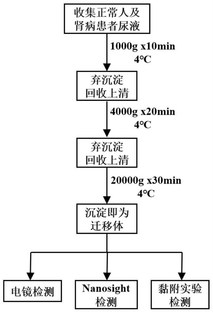
[0035] The urine migration body separation and detection method comprises the following steps (see figure 1 ):

[0036] (1) Collect urine from normal controls and kidney disease patients (FSGS patients) into 50ml centrifuge tubes;

[0037] (2) The following steps are all carried out at 4°C. Use a 4°C pre-cooled high-speed centrifuge to centrifuge at 1000g for 10min, discard the precipitate and recover the supernatant, then centrifuge at 4000g for 20min, discard the precipitate and recover the supernatant, then centrifuge at 20000g for 30min, and the precipitate is The isolated migratory body;

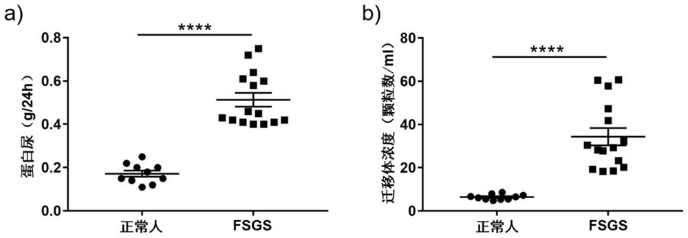
[0038] (3) Electron microscope detection, Nanosight particle size detection and adhesion test were carried out on the separated urine migration body; the results of urine migration body detection of normal people and patients with kidney disease were compared and analyzed.

Embodiment 2

[0040] The specific steps of the detection method are as follows:

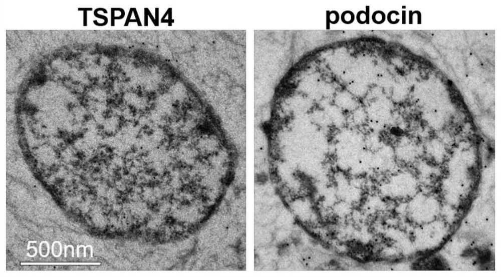
[0041] Immunoelectron microscope detection: (1) The separated urine migratory bodies were fixed overnight at 4°C with 2.5% glutaraldehyde. (2) Rinse with PBS at 4°C. (3) Fix with 1% osmium tetroxide at 4°C. (4) Use ethanol and acetone with concentration gradients for dehydration at 4°C, respectively. (5) Use acetone and resin to impregnate. (6) Use pure resin to embed overnight at 60°C. (7) Ultra-thin slices, nickel net fishing slices. 1% NaIO 4 (ready to use) or 1% H 2 o 2 Etched for 10 minutes, washed 3 times with water, and blocked with electron microscope diluent (1%BSA-PBS) for 15-20 minutes. (8) Incubate primary antibody for 2 hours at room temperature, wash with PBS, incubate gold-labeled secondary antibody for 2 hours at room temperature, wash with PBS, ddH 2 O wash once. (9) Staining, transmission electron microscope observation and photographing.

[0042] Nanosight particle size detection: (...


PUM

PropertyMeasurementUnit
particle diameteraaaaaaaaaa

Abstract

The invention belongs to the technical field of biology, and relates to an application of a urine podocyte-derived migration body in predicting early injury of glomerular podocytes. The migration can be used for predicting the early injury of the glomerular podocytes, prompts a patient to detect other indexes in time, and becomes an effective means for predicting the early injury of the glomerular podocytes.

Description

technical field [0001] The invention belongs to the field of biotechnology, and relates to the application of migratory bodies as biomarkers in the preparation of drugs for predicting early injury of glomerular podocytes. Background technique [0002] Podocyte disease is the main cause of most progressive kidney diseases eventually leading to end-stage renal disease (ESRD). Podocytes control the permeability of glomeruli with their complex morphology. The development of proteinuria is usually associated with podocyte damage. Examples include loss of podocyte foot processes and loss of cells. Since podocytes are terminally differentiated cells and lack capacity for renewal, detection of early podocyte injury is critical for successful treatment of glomerular diseases. [0003] At present, the commonly used diagnostic methods for podocyte injury in clinic include histopathological diagnosis, such as renal puncture, urine protein detection, etc.; etiological diagnosis, such as...

Claims


Application Information

Patent Timeline
Patent Type & Authority Applications(China)
IPC IPC(8): G01N23/04G01N15/02G01N21/64
CPCG01N15/02G01N21/6458G01N23/04
Inventor 曾科李姗刘颖
Owner NANJING UNIV
Why Eureka
Try Eureka