Virus single particle separation method based on nano-micro composite spheres
A separation method and virus particle technology, applied in the directions of microorganism-based methods, viruses/phages, biochemical equipment and methods, etc., can solve the problem of difficulty in obtaining single virus particles, and achieve the effect of low cost
- Summary
- Abstract
- Description
- Claims
- Application Information
AI Technical Summary
Problems solved by technology
Method used
Image

Examples
Embodiment 1
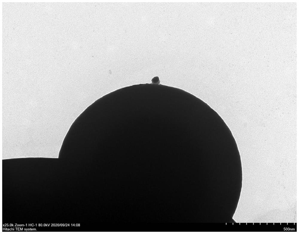
[0031] This implementation discloses a virus single particle separation method based on nano-micro composite spheres. The chip used in this embodiment contains an array of nano-micro composite spheres, wherein the chip is a commercially available 2-inch, 50 μm thick double-sided Polished silicon wafers, the nano-micro composite balls arrayed on the chip are composed of nano-spheres embedded in nano-holes on the surface of the micro-spheres, wherein the micro-spheres are polystyrene micro-spheres with a diameter of 5 μm, the surface is modified by fluorescence, and the interior of the balls is Contains nano-magnetic particles, removes the external magnetic field, has no residual magnetism, and has no virus adsorption capacity. The nanospheres are silica nanospheres with a diameter of 1300nm. The surface is modified by amination and has virus adsorption capacity.
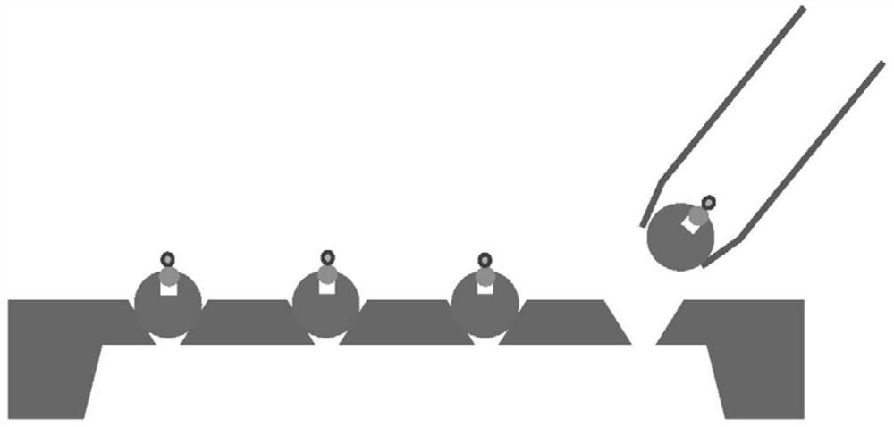
[0032] Single viral particles were isolated using the micropipette method as follows:
[0033] Taking GVE2 phage as...
Embodiment 2
[0036] This implementation discloses a virus single-particle separation method based on nano-micro composite spheres. The chip containing the array of nano-micro composite spheres used in this embodiment, wherein, the chip is commercially available with a thickness of 3 mm and an average filter pore size of 0.45 mm. - 0.8 μm porous ceramics, the nano-micro composite spheres arrayed on the chip are composed of nanospheres embedded in the nanopores on the surface of the microspheres, wherein the microspheres are polystyrene microspheres with a diameter of 2 μm, and the surface is modified by fluorescence , The inside of the ball contains nano-magnetic particles, there is no residual magnetism after removing the external magnetic field, and it does not have the ability to adsorb viruses. The nanospheres are silica nanospheres with a diameter of 100nm. The surface is modified by amination and has virus adsorption ability.
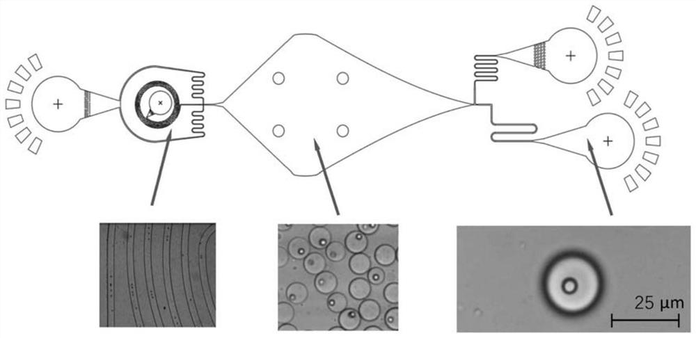
[0037] A droplet microfluidic method is used to separate a...
Embodiment 3
[0040] This implementation discloses a virus single-particle separation method based on nano-micro composite spheres. The chip containing the array of nano-micro composite spheres used in this embodiment, wherein, the chip is commercially available with a thickness of 3 mm and an average filter pore size of 0.45 mm. - 0.8 μm porous ceramics, the nano-micro composite spheres arrayed on the chip are composed of nanospheres embedded in the nanopores on the surface of the microspheres, wherein the microspheres are polystyrene microspheres with a diameter of 2 μm, and the surface is modified by fluorescence , The inside of the ball contains nano-magnetic particles, there is no residual magnetism after removing the external magnetic field, and it does not have the ability to adsorb viruses. The nanospheres are silica nanospheres with a diameter of 100nm. The surface is modified by amination and has virus adsorption ability.
[0041] Individual composite pellets were separated using t...
PUM
| Property | Measurement | Unit |
|---|---|---|
| Diameter | aaaaa | aaaaa |
| Diameter | aaaaa | aaaaa |
| Diameter | aaaaa | aaaaa |
Abstract
Description
Claims
Application Information
 - R&D
- Intellectual Property
- Life Sciences
- Materials
- Tech Scout
- Unparalleled Data Quality
- Higher Quality Content
- 60% Fewer Hallucinations
Browse by: Latest US Patents, China's latest patents, Technical Efficacy Thesaurus, Application Domain, Technology Topic, Popular Technical Reports.
© 2025 PatSnap. All rights reserved.Legal|Privacy policy|Modern Slavery Act Transparency Statement|Sitemap|About US| Contact US: help@patsnap.com