Immunohistochemical combined elastic fiber multi-staining kit, staining method and application

An elastic fiber and immunohistochemical technology, applied in the field of clinical pathology detection, can solve the problems of unsatisfactory specimen quality and quantity, high requirements for pathological specimens, cumbersome steps in the production process, etc., achieve clear and intuitive color contrast, and improve detection Efficiency, ease of diagnosis and analysis

Pending Publication Date: 2021-06-22
河南赛诺特生物技术有限公司
View PDF0 Cites 3 Cited by
  • Summary
  • Abstract
  • Description
  • Claims
  • Application Information

AI Technical Summary

Problems solved by technology

This not only results in cumbersome steps in the production process and low efficiency of the diagnostic process, but also has high requirements for pathological specimens, especially for lung cancer puncture tissue, because lung puncture examination is an invasive examination, and the skills of puncture doctors are different and the patient's Due to individual differences, the number of puncture tissue samples obtained is often small, and the quality and quantity of specimens are often unsatisfactory. It is difficult to obtain a sufficient number of sections for routine HE staining, immunohistochemical staining, Fish fluorescence in situ hybridization staining, or special staining. Item detection

Method used


Image

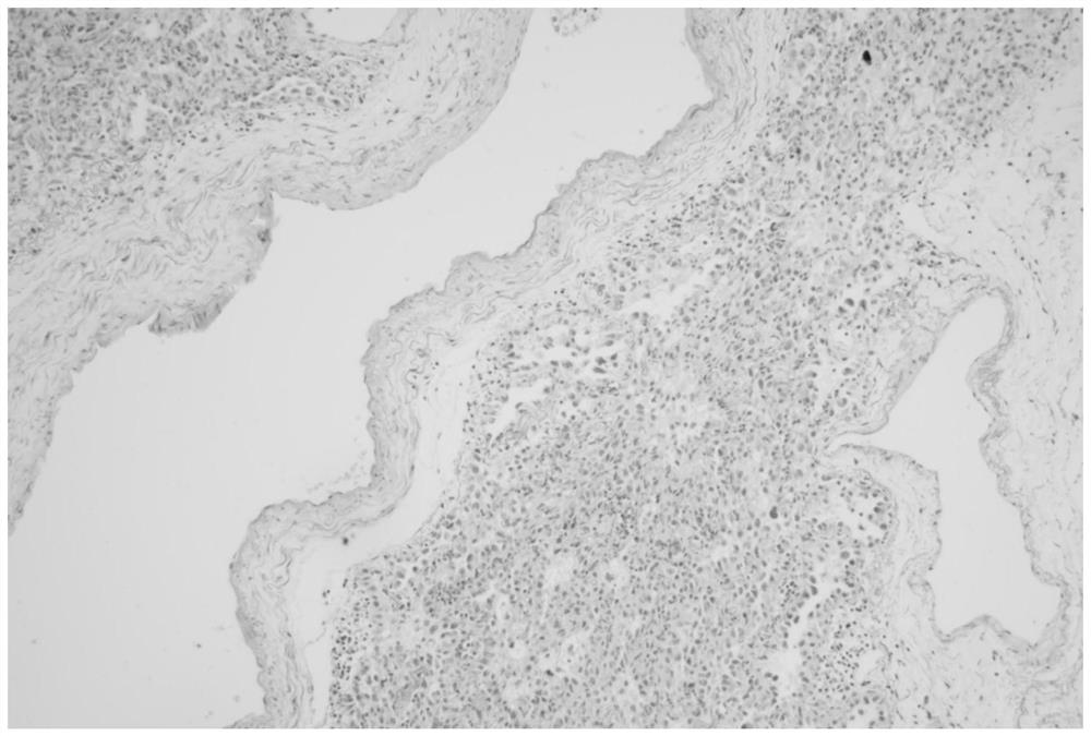
  • Immunohistochemical combined elastic fiber multi-staining kit, staining method and application
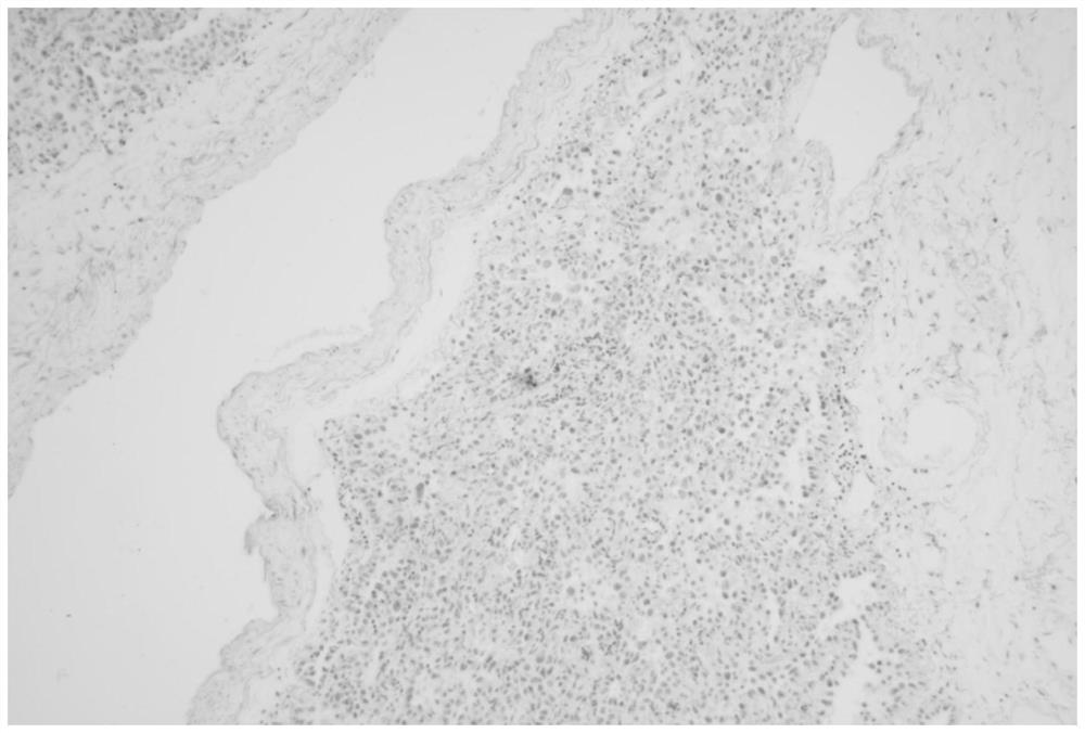
  • Immunohistochemical combined elastic fiber multi-staining kit, staining method and application
  • Immunohistochemical combined elastic fiber multi-staining kit, staining method and application

Examples


Embodiment 1

[0070] Example 1 Immunohistochemical combined elastic fiber multi-dye kit 1

[0071] This embodiment provides an immunohistochemical combined elastic fiber multi-diery kit, including the following components:

[0072] 1) Antigen repair liquid: EDTA repair fluid (pH9.0)

[0073] 2) Victoria blue staining solution: It is mainly composed of Victoria blue, inter-amphibenzol, and iron trichloride.

[0074] 3) Peroxidase blockage is purchased from Henan Sainte Biotechnology Co., Ltd., the item number RS3001.

[0075] 4) Anti-reagent: The Anti-reagent is a mixed antibody composed of antibody A and an antibody B, which is an anti-TTF-1 mouse monoclonal antibody (cloning 8G7G3 / 1, Henan Sainoke biotechnology Ltd., Item No. CTM-0261), the antibody B is anti-P40 rabbit monoclonal antibody (cloning number C3B4, Henan Sainte Biotechnology Co., Ltd., Item No. CPM-0133);

[0076] 5) Second antibody A: HRP enzyme labeled sheep anti-rat polymer, purchased from Henan Sainte Biotechnology Co., Ltd....

Embodiment 2

[0088] Example 2 Immunohistochemical combined elastic fiber multi-diery kit 2

[0089] This embodiment provides an immunohistochemical combined elastic fiber multi-diery kit, including the following components:

[0090] 1) Antigen repair liquid: EDTA repair fluid (pH9.0)

[0091] 2) Victoria blue staining solution: It is mainly composed of Victoria blue, inter-amphibenzol, ferricide, and formula 1, refer to Example 1.

[0092] 3) Peroxidase Closure: Purchased from Henan Sainte Biotechnology Co., Ltd., the item number RS3001.

[0093] 4) Anti-reagent: The antibody agent is a mixed antibody composed of antibody A and antibody B, which is an anti-ER mouse monoclonal antibody (cloning C6H7, Henan Sainte Biotechnology Co., Ltd., Item Number CEM-0081), the antibody B is anti-HER2 rabbit monoclonal antibody (rabbit cloned EP3, Henan Sainte Biotechnology Co., Ltd., Item No. CCR-0743);

[0094] 5) Second antibody A: HRP enzyme labeled sheep anti-rat polymer, purchased from Henan Sainte Bio...

Embodiment 3

[0133] Example 3 Immunohistochemical combined elastic fiber poly-dyeing method 1

[0134] This contravention provides a poly-immunohistochemical combined elastic fiber multi-dyeing method: After the tissue sliced ​​dewaxing, the immunohistochemical high pressure repair, no potassium oxidation of permanganate, advanced elastic fiber staining, and then immunohistochemical double dyeing , Including the following steps:

[0135] 1) Dewaxing and hydration: from xylene to anhydrous ethanol, then soaked to 95%, 85%, 75% gradient ethanol soaked, to hydration.

[0136] 2) antigen repair;

[0137] 3) Elastic fiber staining: 95% ethanol is washed slightly, and Victoria blue staining is incubated for 1 hour;

[0138] 4) Differentiation: 95% ethanol is quickly dipped to there is no excess drying solution;

[0139] 5) Purify water soaked, rinse;

[0140] 6) Add 100 μl of peroxidase enzyme, incubate for 5 min at room temperature; wash liquid;

[0141] 7) Add 100 μL of an anti-reagent, incubate a...


PUM


Abstract

The invention relates to an immunohistochemical combined elastic fiber multi-staining kit, a staining method and application. The kit provided by the invention comprises the following components: an antigen repair solution, a Victoria blue dye solution, a peroxidase blocking agent, a primary antibody reagent, a secondary antibody reagent A, a secondary antibody reagent B, a DAB chromogenic substrate, a DAB chromogenic buffer solution, an HRP-Blue substrate, an HRP-Blue substrate buffer solution and a hematoxylin counterstaining solution. The primary antibody reagent is a mixed antibody mainly composed of an antibody A and an antibody B, the antibody A is a mouse monoclonal antibody, and the antibody B is a rabbit monoclonal antibody; the second antibody reagent A is an HRP (horse radish peroxidase) labeled goat anti-mouse polymer; the second antibody reagent B is an HRP (horse radish peroxidase) labeled goat anti-rabbit polymer. The invention also provides a specific dyeing method. According to the kit and the dyeing method, immunohistochemical double dyeing and elastic fiber dyeing are organically combined for the first time, the dyeing procedure is simplified, the detection efficiency is improved, and the demand for tissue samples is remarkably reduced.

Description

Technical field [0001] The present invention belongs to the technical field of clinical pathological detection, and more particularly to an immunohistochemical combined elastic fiber multimodal kit, dyeing method and application. Background technique [0002] Immunohistochemistry, IHC is also known as immunocytochemical dyeing, referred to as immunohistochemistry, is the most important platform technique for pathological diagnosis, differential diagnosis, tumor-specific molecular target screening, identification. In recent years, in the diagnosis and differential diagnosis of malignant tumors; the primary part of the metastatic malignant tumor; further pathological division for a type of tumor; research on immunohistochemistry to use a variety of signs to study the diagnosis of soft tissue tumors is indispensable It is discovered that a small transfer stove will help the clinical treatment plan, including the determination of the surgical range; it has been widely used in the sel...

Claims


Application Information

Patent Timeline
Patent Type & Authority Applications(China)
IPC IPC(8): G01N1/31G01N33/532G01N33/535G01N33/577
CPCG01N1/31G01N33/532G01N33/535G01N33/577
Inventor 梁永波李莉陈玲李三华刘畅牛银银齐华刘文弟
Owner 河南赛诺特生物技术有限公司
Why Eureka
Try Eureka