T cell proliferation method and application thereof

A cell and cell suspension technology, applied in the field of 3D bioprinting T cell proliferation, can solve the problems of high degree of cell exhaustion, high cost of CAR-T cell preparation, and difficulty in obtaining T cells, and achieve the effect of improving survival rate.

Pending Publication Date: 2021-06-25
华东数字医学工程研究院
View PDF6 Cites 0 Cited by
  • Summary
  • Abstract
  • Description
  • Claims
  • Application Information

AI Technical Summary

Problems solved by technology

There are still many problems with this therapy. For example, it is difficult to obtain a sufficient amount of T cells in some young or elderly patients and patients with high tumor burden, and the preparation cost of CAR-T cells is high, and the production time is long, so it cannot be shortened. A sufficient amount of T cells can be obtained by culturing and proliferating within a certain period of time, and the degree of cell exhaustion and cell necrosis and apoptosis are relatively high

Method used


Image

  • T cell proliferation method and application thereof
  • T cell proliferation method and application thereof
  • T cell proliferation method and application thereof

Examples


Embodiment 1

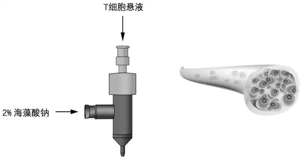
[0088] Example 1: Three-dimensional coaxial printing to obtain T cells

[0089] experimental method:

[0090] Take 20mL of healthy human blood, add it to a heparin anticoagulant tube, add 20mL of PBS to dilute, slowly add 10mL of Ficoll separation solution, and use a centrifuge to centrifuge at 2000r / min for 20 minutes; absorb the milky white layer after centrifugation, that is, PBMC, and then add an appropriate amount of PBS to wash , and finally add an appropriate amount of PBS for counting.

[0091] Next, wash the PBMC with an appropriate amount of MACS buffer, and continue to use MACS buffer to resuspend, add CD3 immunomagnetic beads (20μL / 10 7 PBMC) mix well, and incubate at 4°C for 15 minutes; wash the cells again with MACS buffer, resuspend and add to the washed MS separation column, the cells that flow out are CD3-T cells, wash the isolate 3 times with MACS buffer, and finally add 1mL of cells were eluted, and the eluate was CD3+T cells, which were counted and resusp...

Embodiment 2

[0098] Example 2: Obtaining T cells in suspension culture

[0099] The processing before printing is the same as that in Embodiment 1. T cells were divided into 1 x 10 6 / ml density inoculated in T25 culture flasks, and the solution was changed every three days.

Embodiment 3

[0100] Example 3: Comparison of proliferation ability of T cells obtained by three-dimensional coaxial printing and T cells obtained by suspension culture

[0101] experimental method:

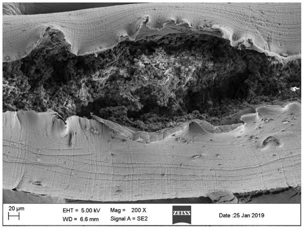
[0102] In order to evaluate the proliferation ability of T cells in coaxial bioprinted T cell-loaded hydrogel fibers, we used a flow cytometer (Beckman, CytoFlex flow cytometer) to analyze the fluorescence intensity of CFSE in T cells (the fluorescence intensity was halved, the cells Division once), which represents the division algebra of T cells. On the 3rd, 7th, and 10th day after printing, we used citric acid-EDTA solution to dissolve the sodium alginate shell of the coaxial fiber, and collected quantitative cells to detect the fluorescence intensity of CFSE.

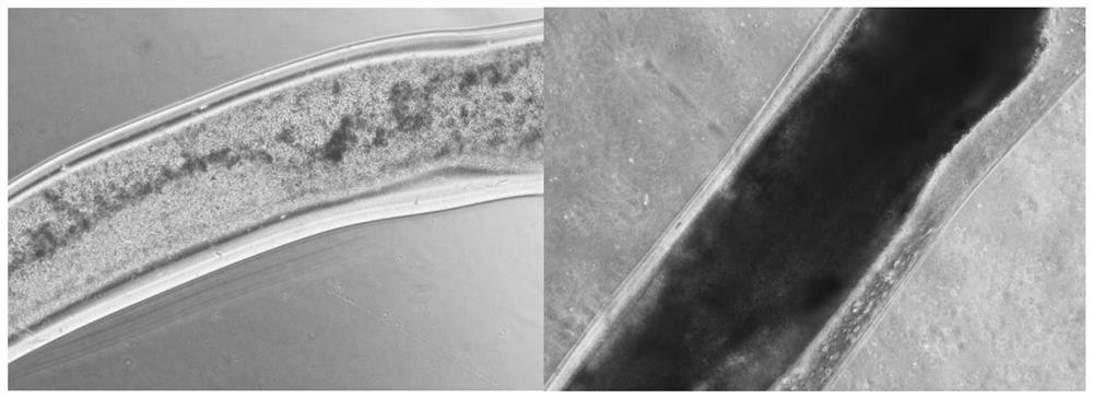
[0103] Experimental results:

[0104] Experimental results such as image 3 and Figure 4 shown.

[0105] Such as image 3 As shown, by observing the micrographs of the 3D coaxially printed T cells on the 3rd day (left) and ...


PUM


Abstract

The invention relates to a T cell proliferation method and an application thereof, and belongs to the field of biomedicine. The method comprises the following steps: obtaining a cell cellosilk, wherein the cell cellosilk comprises an inner core and a fiber outer layer wrapping the inner core; wherein the inner core is derived from a cell suspension containing T cells, and the fiber outer layer is derived from a high polymer material; and culturing the cell cellosilk to obtain the T cell containing the fiber outer layer. According to the T cell proliferation method provided by the invention, the in-vitro proliferation capability of the T cells is improved, the survival rate of the T cells is increased, a new thought is provided for in-vitro culture of the T cells, and a new cell culture method is provided for adoptive immunotherapy.

Description

technical field [0001] The invention belongs to the field of biotechnology or three-dimensional bioprinting, and specifically relates to a three-dimensional bioprinting T cell proliferation method and its application. Background technique [0002] Coaxial extrusion bioprinting is a novel approach to construct a three-dimensional microenvironment that simulates cells. Coaxial channels can have multiple layers, creating thread-like structures composed of multiple materials and cells. Based on coaxial printing technology, a shell-core structure for carrying cells in tissue engineering, drug screening and in vitro model research of tissues and organs was constructed, so that the cells were in a three-dimensional environment, and the corresponding microstructure was provided by the extracellular matrix material forming the "shell". Environment, so that the cells are in a microenvironment similar to that in the body, which is conducive to the biological functions of the cells. ...

Claims


Application Information

Patent Timeline
IPC IPC(8): C12N5/0783A61K39/00A61P35/00
CPCC12N5/0636A61K39/001111A61P35/00C12N2513/00A61K2039/515
Inventor 金治中韩帅龙小燕
Owner 华东数字医学工程研究院
Why Eureka
Try Eureka