Cell image segmentation method based on anti-background differencing

A background subtraction and image segmentation technology, which is applied in the field of cell evaluation and cell image processing, can solve the problems that are not suitable for rapid and large-scale analysis of cell data, over-segmentation, and not suitable for cell segmentation, etc., to achieve high segmentation efficiency, eliminate halo, The effect of suppressing interfering factors

Pending Publication Date: 2021-07-23
NINGBO UNIV +1
View PDF10 Cites 2 Cited by
  • Summary
  • Abstract
  • Description
  • Claims
  • Application Information

AI Technical Summary

Problems solved by technology

Cell segmentation is affected by differences between subcellular structures, and cell image intensity gradients are small inside the cell and large around the cell membrane, often leading to over-segmentation by eg marker-controlled watershed segmentation algorithms
Cell segmentation is possible using machine learning-based methods, such as instance segmentation, random forest, U-Net model, etc., but may not be suitable for rapid large-scale analysis of cell data due to time-consuming manual labeling of cell data and model training
In addition, since the morphological characteristics of MSCs are constantly changing, methods using morphological models may not be suitable for cell segmentation

Method used


Image

  • Cell image segmentation method based on anti-background differencing
  • Cell image segmentation method based on anti-background differencing
  • Cell image segmentation method based on anti-background differencing

Examples


Embodiment 1

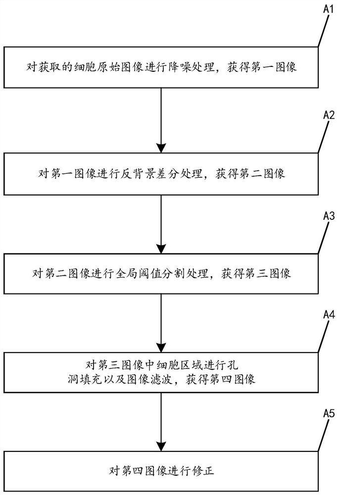
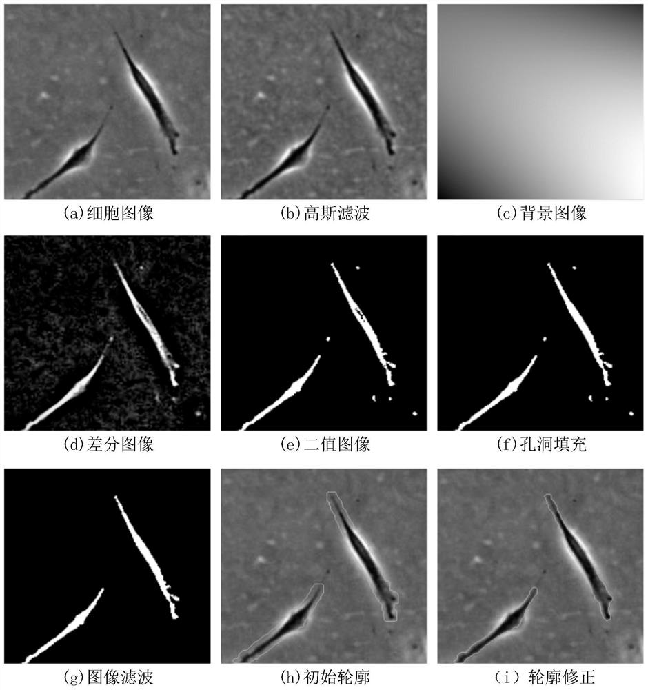
[0029] figure 1 The flow chart of the cell image segmentation method of inverse background difference according to the embodiment of the present invention is given, such as figure 1 As shown, the cell image segmentation method of inverse background difference comprises the following steps:

[0030] (A1) noise reduction processing is performed on the acquired original cell image to obtain a first image;

[0031] (A2) performing inverse background difference processing on the first image to obtain a second image;

[0032] (A3) global threshold segmentation processing is performed on the second image to obtain a third image;

[0033] (A4) Performing hole filling and image filtering on the cell region in the third image to obtain a fourth image.

[0034] In order to improve the effect of image segmentation, further, the cell image segmentation method further includes the following steps:

[0035] (A5) Correcting the fourth image, the correction method is: performing morphologi...

Embodiment 2

[0058] An application example of the cell image segmentation method according to the inverse background difference of the embodiment 1 of the present invention in the evaluation of mesenchymal stem cells.

[0059] The cell image segmentation method of inverse background difference according to the embodiment of the present invention includes the following steps:

[0060] (A1) noise reduction processing is performed on the acquired original cell image to obtain a first image;

[0061] The specific method for obtaining the original image of the cell is as follows:

[0062] Cell recovery: add 9mL of complete cell culture medium to a 15mL sterile centrifuge tube, and place it in a 37°C water bath to preheat. Take out the cryovial containing adult bone marrow mesenchymal stem cells, put it in a 37°C water bath, and keep shaking until the contents are thawed. Use 70%-75% alcohol to disinfect the opening and outer wall of the cryopreservation tube and wipe it clean.

[0063] Open ...


PUM


Abstract

The invention provides an anti-background difference-based cell image segmentation method. The method comprises the steps: (A1) carrying out the noise reduction of an obtained original cell image, and obtaining a first image; (A2) performing anti-background difference processing on the first image to obtain a second image; (A3) carrying out global threshold segmentation processing on the second image to obtain a third image; and (A4) carrying out hole filling and image filtering on the cell region in the third image to obtain a fourth image. The method has the advantages of high segmentation precision, high efficiency and the like.

Description

technical field [0001] The present invention relates to cell evaluation and cell image processing methods, in particular to a cell image segmentation method based on inverse background difference. Background technique [0002] Mesenchymal stem cells (MSCs) have multi-directional differentiation potential and immunomodulatory properties, and can differentiate into osteogenic, adipose and chondrogenic cell lines under appropriate induction conditions, and are widely used in various tissue regeneration applications and immune-related Treatment of diseases such as acute and chronic graft-versus-host disease. However, the amount of MSCs isolated from bone marrow and other tissues is very small, which cannot meet the demand of MSCs in research and clinical treatment, so large-scale expansion and culture in vitro is required. Cell preparations that meet quality standards are extremely important for subsequent cell therapy applications. The current use of molecular biology standar...

Claims


Application Information

Patent Timeline
Patent Type & Authority Applications(China)
IPC IPC(8): G06T7/00G06T7/136G06T5/00
CPCG06T7/0012G06T7/136G06T5/002G06T2207/10056G06T2207/20024G06T2207/30004
Inventor 史振志吴京城施露露闻路红胡舜迪
Owner NINGBO UNIV
Why Eureka
Try Eureka