Cell image segmentation method and device, electronic equipment and storage medium

A cell and image technology, applied in the field of image processing, can solve problems such as consuming a lot of time, under-segmentation, and over-segmentation, and achieve the effects of saving manpower and time, improving efficiency and accuracy, and avoiding errors

Pending Publication Date: 2021-07-27
XIAN JIAOTONG LIVERPOOL UNIV
View PDF4 Cites 2 Cited by
  • Summary
  • Abstract
  • Description
  • Claims
  • Application Information

AI Technical Summary

Problems solved by technology

[0003] The traditional method of cell nucleus segmentation is supervised learning using human-labeled data sets, which takes a lot of time to label mask images, and the labeling process varies from person to person, and the obtained data sets have expert bias, so the segmentation effect of this type of method is limited
And it requires a lot of manpower, it is easy to cause over-segmentation or under-segmentation, and the segmentation efficiency and accuracy of cell images are low

Method used


Image

  • Cell image segmentation method and device, electronic equipment and storage medium
  • Cell image segmentation method and device, electronic equipment and storage medium
  • Cell image segmentation method and device, electronic equipment and storage medium

Examples


Embodiment 1

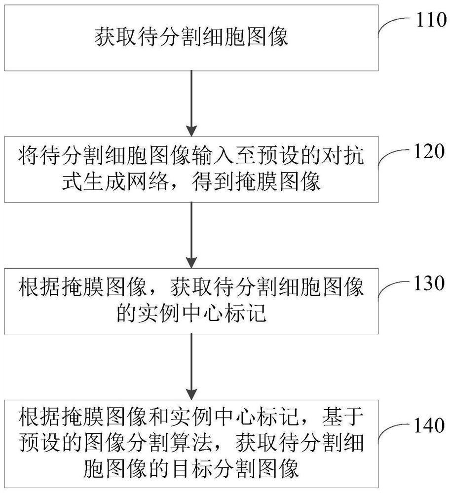
[0024] figure 1 It is a schematic flowchart of a cell image segmentation method provided in Embodiment 1 of the present invention. This embodiment is applicable to the case of cell image segmentation, and the method can be executed by a cell image segmentation device. Such as figure 1 As shown, the method specifically includes the following steps:

[0025] Step 110, acquiring the image of the cell to be segmented.

[0026] Wherein, the image of the cell to be segmented may be a three-dimensional image, for example, it may be a CLSM three-dimensional image of cell culture to be segmented, and the CLSM three-dimensional image is an image taken by a CLSM microscope. The method for acquiring the image of the cell to be segmented may be to acquire a three-dimensional image under a CLSM microscope.

[0027] Step 120, input the image of the cell to be segmented into a preset confrontational generation network to obtain a mask image.

[0028] Wherein, an adversarial generation net...

Embodiment 2

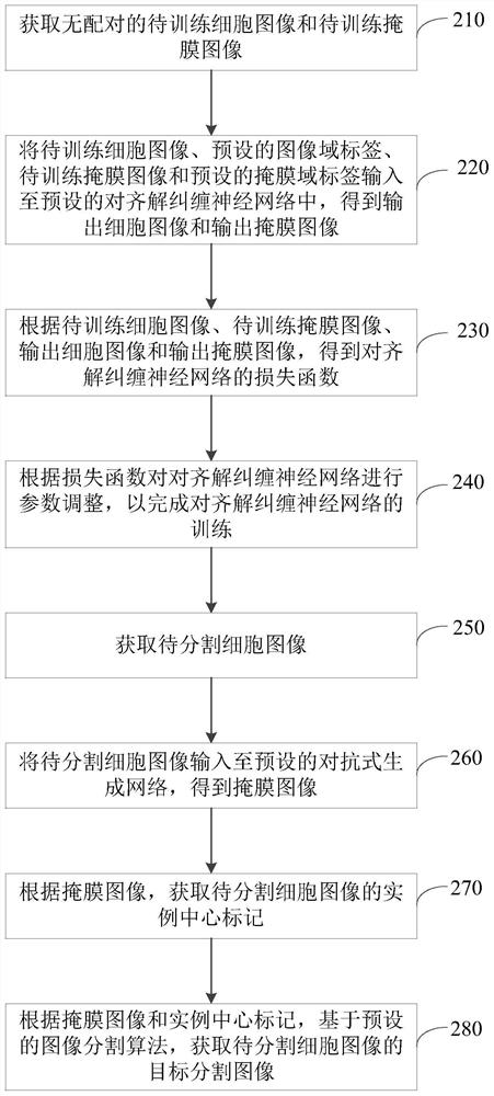
[0044] figure 2 It is a schematic flowchart of a cell image segmentation method provided in Embodiment 2 of the present invention. This embodiment is further optimized on the basis of the above embodiments, and the method can be executed by a cell image segmentation device. Such as figure 2 As shown, the method specifically includes the following steps:

[0045] Step 210, acquiring unpaired cell images to be trained and mask images to be trained.

[0046] Wherein, the image of the cell to be trained is a pre-acquired sample image, and the sample image forms an image domain. There can be multiple cells in a cell image to be trained. The staff roughly estimates the number and size of nuclei in the image domain, and randomly generates a mask image to be trained to form a mask domain according to the estimated size and number of nuclei. For example, a certain number of ellipsoids with a certain size range can be randomly generated, and the ellipsoids can be randomly placed in...

Embodiment 3

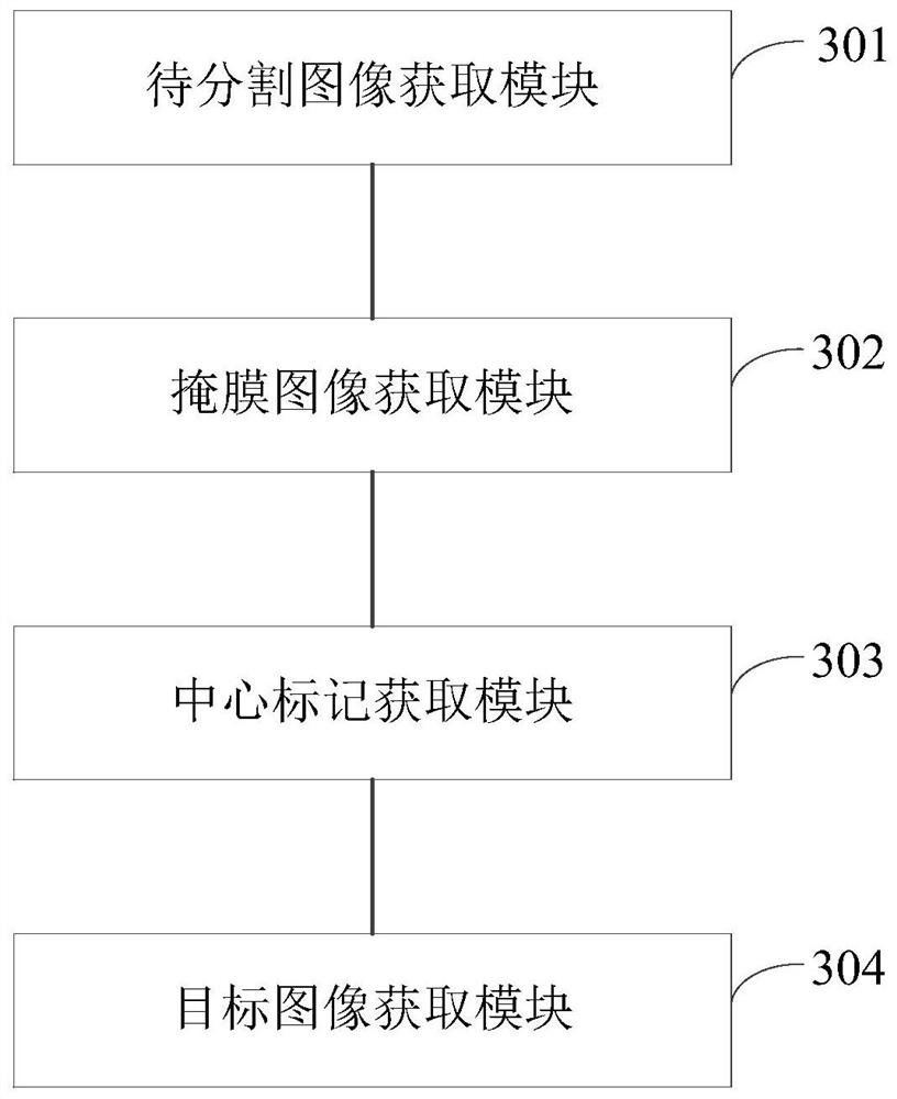
[0071] image 3 It is a structural block diagram of a cell image segmentation device provided in Embodiment 3 of the present invention, which can execute the cell image segmentation method provided in any embodiment of the present invention, and has corresponding functional modules and beneficial effects for executing the method. Such as image 3 As shown, the device specifically includes:

[0072] An image to be segmented acquisition module 301, configured to acquire an image of a cell to be segmented;

[0073] A mask image acquisition module 302, configured to input the image of the cell to be segmented into a preset confrontational generation network to obtain a mask image;

[0074] A central marker acquisition module 303, configured to acquire an instance central marker of the cell image to be segmented according to the mask image;

[0075] The target image acquisition module 304 is configured to acquire the target segmented image of the cell to be segmented based on th...


PUM


Abstract

The embodiment of the invention discloses a cell image segmentation method and device, electronic equipment and a storage medium. The method comprises the following steps: acquiring a to-be-segmented cell image; inputting the to-be-segmented cell image into a preset generative adversarial network to obtain a mask image; obtaining an instance center mark of the to-be-segmented cell image according to the mask image; and according to the mask image and the instance center mark, based on a preset image segmentation algorithm, obtaining a target segmentation image of the to-be-segmented cell. The to-be-segmented cell image is input into the preset generative adversarial network model, the mask image of the to-be-segmented cell image is automatically generated, and the cell nucleus of the to-be-segmented cell image is labeled, so that cell nucleus segmentation is performed on the to-be-segmented cell image, manual cell nucleus labeling on the to-be-segmented cell image is avoided, manual operation steps are reduced, and the cell segmentation efficiency and precision are improved.

Description

technical field [0001] Embodiments of the present invention relate to image processing technology, and in particular to a cell image segmentation method, device, electronic equipment, and storage medium. Background technique [0002] CLSM (Confocal laser scanning microscope) images contain a large amount of information on cell and tissue structures, and are widely used in evaluating three-dimensional cell cultures based on biological scaffolds. Since the morphology, distribution, nucleoplasmic ratio, and structure of the nucleus are the basis for studying subcellular information, the segmentation of the nucleus in 3D cell culture is an important task in the study and analysis of 3D cell culture. [0003] The traditional method of cell nucleus segmentation is supervised learning using human-labeled data sets, which takes a lot of time to label mask images, and the labeling process varies from person to person, and the obtained data sets have expert bias, so the segmentation e...

Claims


Application Information

Patent Timeline
Patent Type & Authority Applications(China)
IPC IPC(8): G06T7/11G06T7/136G06N3/04G06N3/08
CPCG06T7/11G06T7/136G06N3/04G06N3/088G06T2207/10056G06T2207/20081G06T2207/30024
Inventor 黄开竹姚凯孙捷
Owner XIAN JIAOTONG LIVERPOOL UNIV
Why Eureka
Try Eureka