Cell analysis method based on cell nucleus DNA analysis, computer equipment and storage medium

A cell nucleus and cell technology, applied in the field of cell analysis, can solve the problems of low accuracy of sample image data processing, influence of the accuracy of analysis results, missed diagnosis and misdiagnosis by medical personnel, etc., to facilitate analysis and observation, and improve sensitivity and accuracy. efficiency, reducing workload

Active Publication Date: 2021-07-27
ZHENGZHOU ZHONGPU MEDICAL INSTR CO LTD
View PDF12 Cites 2 Cited by
  • Summary
  • Abstract
  • Description
  • Claims
  • Application Information

AI Technical Summary

Problems solved by technology

[0003] With the development of AI technology, more and more rely on AI technology in cell analysis. However, the existing AI technology for biological samples The following problems still commonly exist in the analysis work: the accuracy rate of sample image data processing is not high, which is easy to cause analysis errors; most of the existing biological sample analysis results only output positive and negative output results, and biological samples in the intermediate state belong to The analysis blind spot, therefore, has a certain impact on the accuracy of the analysis results, and is prone to problems such as missed diagnosis and misdiagnosis by medical personnel

Method used


Image

  • Cell analysis method based on cell nucleus DNA analysis, computer equipment and storage medium
  • Cell analysis method based on cell nucleus DNA analysis, computer equipment and storage medium
  • Cell analysis method based on cell nucleus DNA analysis, computer equipment and storage medium

Examples


Embodiment 1

[0032] The cell analysis method based on nuclear DNA analysis of the present invention comprises the following steps:

[0033] S1: obtaining an image of an isolated biological sample;

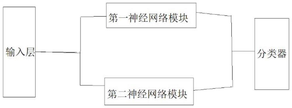
[0034] S2: Analyzing the first appearance of the organism in the isolated biological sample in the image to determine the physical or pathological properties of the isolated biological sample;

[0035] S3: Analyzing the second appearance of the organism in the isolated biological sample in the image to determine the physical or pathological properties of the isolated biological sample;

[0036] S4: Obtain the cell analysis result of the isolated biological sample.

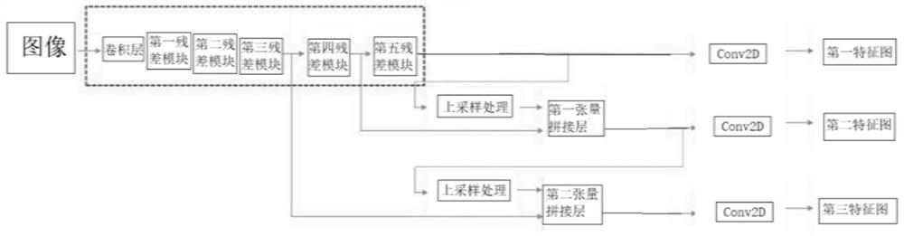
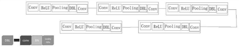
[0037] The image in step S1 can be an image of an original unstained biological sample, or a dyed biological sample.

[0038] The staining method of the biological sample can adopt any staining method in the prior art, such as wilt-Nissl acid-fast staining (mycobacterium tuberculosis is stained red under the microscope, other non-tu...

Embodiment 2

[0066] This embodiment provides a computer device, which includes: a processor; a memory for storing a computer program; when the computer program is executed by the processor, the processor implements the steps of the detection method described in Embodiment 1 .

[0067] Wherein, the processor can be a central processing unit (CPU) or a field programmable logic array (FPGA) or a single-chip microcomputer (MCU) or a digital signal processor (DSP) or an application-specific integrated circuit (ASIC), etc., which have data processing capabilities and / or programs A device capable of performing logic operations. One or more processors may be configured to execute the above-mentioned detection method at the same time as a parallel computing processor group, or configured to use some processors to execute some steps in the above-mentioned detection method, and some processors to execute other steps in the above-mentioned detection method. some steps etc. Computer instructions incl...

Embodiment 3

[0074] This embodiment is a computer-readable storage medium, on which a computer program is stored, and when the computer program is run by a processor, the steps of the method described in the first embodiment are executed.


PUM


Abstract

The invention discloses a cell analysis method based on cell nucleus DNA analysis, computer equipment and a storage medium. The cell analysis method comprises a first appearance form judgment step and a second appearance form judgment step; the first appearance form judgment step is realized by obtaining a parameter of the area size of a cell nucleus, defining the parameter as a DNA parameter 1, respectively presetting a threshold value A, a threshold value B and a threshold value C for the DNA parameter 1, and dividing the DNA parameter 1 into four numerical ranges, thereby dividing the cells to be analyzed into four types: abnormal cells, suspicious abnormal cells, highly suspicious abnormal cells and positive abnormal hyperplasia cells; and further calculating and judging the coincidence probability of the four types of cells by utilizing a second appearance form judgment step. Therefore, by utilizing the method disclosed by the invention, the speed of cell analysis results can be increased, the workload of medical personnel can be reduced, the accuracy of the output analysis results can be ensured, and the problems of missed diagnosis, misdiagnosis and the like can be avoided.

Description

technical field [0001] The invention relates to a cell analysis method, in particular to a cell analysis method based on nuclear DNA analysis, computer equipment and a storage medium. Background technique [0002] Many lesions in organisms can cause abnormalities in normal cells. Therefore, in clinical practice, it is often necessary to use the results of microscopic examination of biological samples to determine whether lesions occur in tissues or organisms. [0003] With the development of AI technology, more and more rely on AI technology in cell analysis work. However, the existing analysis work of biological samples using AI technology still generally has the following problems: the accuracy rate of sample image data processing It is not high, and it is easy to cause analysis errors; most of the existing biological sample analysis results only output positive and negative output results, and biological samples in the intermediate state are analysis blind spots, so it ha...

Claims


Application Information

Patent Timeline
Patent Type & Authority Applications(China)
IPC IPC(8): G16B20/30G06K9/62G06N3/04G06N3/08
CPCG16B20/30G06N3/08G06N3/045G06F18/23213G06F18/2415
Inventor 詹晓春詹斯喻付凤霞张海燕王静孙雷潘建华谭云洪孙国清
Owner ZHENGZHOU ZHONGPU MEDICAL INSTR CO LTD
Why Eureka
Try Eureka