Method for separating and culturing mouse small intestine organoid capable of peristalsis in vitro

A technology of organoids and small intestines, which is applied in the field of cultivating peristaltic mouse small intestine organoids and in vitro separation, which can solve the problems of complicated operation, difficult cultivation, and inability to observe for a long time, and achieve the effect of simple operation method

Inactive Publication Date: 2021-08-03
宁波思成生物科技有限公司
View PDF1 Cites 1 Cited by
  • Summary
  • Abstract
  • Description
  • Claims
  • Application Information

AI Technical Summary

Problems solved by technology

[0005] The currently used small intestinal smooth muscle in vitro experiments mainly use small intestinal smooth muscle tissue (and surrounding tissues) for drug sensitivity tests, etc. The disadvantage is that long-term observation or drug sensitivity tests cannot be performed, and it requires special treatment such as matrigel coating, which is difficult to cultivate and difficult to operate. complex

Method used


Image

  • Method for separating and culturing mouse small intestine organoid capable of peristalsis in vitro
  • Method for separating and culturing mouse small intestine organoid capable of peristalsis in vitro
  • Method for separating and culturing mouse small intestine organoid capable of peristalsis in vitro

Examples


Embodiment 1

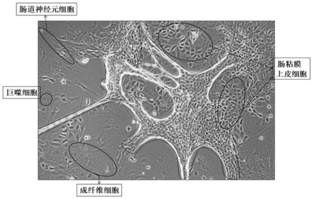
[0039] 1. Separation method

[0040] Step 1: Take three c57BL mice one week after birth, kill them by cervical dislocation, and sterilize them by immersing them in 70% alcohol for 1 min;

[0041] Step 2: Dissect the mouse in the ultra-clean workbench, take out all the large intestine and small intestine, put them in a 100mm Petri dish filled with PBS, and remove the large intestine;

[0042] Step 3: Using ophthalmic scissors, cut the small intestine longitudinally along the small intestine;

[0043] Step 4: Use the curved convex side of the curved ophthalmic forceps to gently scrape off the residues inside the small intestine and most of the small intestinal mucosal epithelium;

[0044] Step 5: Wash 2-3 times with 1xPBS buffer. After removing the washing supernatant, use ophthalmic scissors to cut the small intestine tissue into a size less than 1mm 3 Tissue fragments, and transferred to a 50mL centrifuge tube;

[0045] Step 6: After washing twice with 1xPBS, centrifuge wi...

Embodiment 2

[0063] 1. Separation method

[0064] Step 1: Take three c57BL mice one week after birth, kill them by cervical dislocation, and sterilize them by immersing them in 70% alcohol for 1 min;

[0065] Step 2: Dissect the mouse in the ultra-clean workbench, take out all the large intestine and small intestine, put them in a 100mm Petri dish filled with PBS, and remove the large intestine;

[0066] Step 3: Using ophthalmic scissors, cut the small intestine longitudinally along the small intestine;

[0067] Step 4: Use the curved convex side of the curved ophthalmic forceps to gently scrape off the residues inside the small intestine and most of the small intestinal mucosal epithelium;

[0068] Step 5: Wash 2-3 times with 1xPBS buffer. After removing the washing supernatant, use ophthalmic scissors to cut the small intestine tissue into a size less than 1mm 3 Tissue fragments, and transferred to a 50mL centrifuge tube;

[0069] Step 6: After washing twice with 1xPBS, centrifuge wi...

Embodiment 3

[0087] 1. Separation method

[0088] Step 1: Take three c57BL mice one week after birth, kill them by cervical dislocation, and sterilize them by immersing them in 70% alcohol for 1 min;

[0089] Step 2: Dissect the mouse in the ultra-clean workbench, take out all the large intestine and small intestine, put them in a 100mm Petri dish filled with PBS, and remove the large intestine;

[0090] Step 3: Using ophthalmic scissors, cut the small intestine longitudinally along the small intestine;

[0091] Step 4: Use the curved convex side of the curved ophthalmic forceps to gently scrape off the residues inside the small intestine and most of the small intestinal mucosal epithelium;

[0092] Step 5: Wash 2-3 times with 1xPBS buffer. After removing the washing supernatant, use ophthalmic scissors to cut the small intestine tissue into a size less than 1mm 3 Tissue fragments, and transferred to a 50mL centrifuge tube;

[0093] Step 6: After washing twice with 1xPBS, centrifuge wi...


PUM


Abstract

The invention discloses a method for separating and culturing mouse small intestine organoid capable of peristalsis in vitro, and belongs to the technical field of biological medicine, and the method comprises the following steps: carrying out cell separation and cell culture on mouse small intestines. Different growth periods of animals can be simulated for observation or various drug experiments and toxicity experiments, so that mechanism research on organisms, basic research on diseases such as etiology and pathogenesis, new drug screening, preclinical evaluation of precise treatment of intestinal diseases, relation research with intestinal microorganisms, disease prediction, prevention research and the like are further realized. The method does not need special treatment such as matrigel coating, only needs a common culture bottle and a common culture medium for culture, and is simple in operation method.

Description

technical field [0001] The invention relates to the technical field of biomedicine, in particular to a method for isolating and cultivating peristaltic mouse small intestine organoids in vitro. Background technique [0002] The study of planar culture (2D culture) using cell lines has a huge amount of research data as a reference, but it has limitations in the mechanism research of tissues, organs and even living organisms, as well as in drug development and clinical application, which are greatly different from those in vivo. . In a physiological environment, a variety of cells need to "communicate" with each other and be affected by the cell matrix (ECM), and complex biological functions such as gene transcription, receptor expression, cell migration, and programmed death of different cells occur. [0003] Organoids are in vitro three-dimensional cultures composed of multi-lineage cells, driven by stem cells and constructed in a self-organized manner, which can simulate t...

Claims


Application Information

Patent Timeline
Patent Type & Authority Applications(China)
IPC IPC(8): C12N5/071C12N5/077
CPCC12N5/0661C12N5/0625C12N5/0697C12N2509/00C12N2509/10
Inventor 刘广成叶玲玲王坚强潘魁魁金启凡
Owner 宁波思成生物科技有限公司
Why Eureka
Try Eureka