Method for identifying tumor boundary by carrying out molecular spectral imaging on cancer tissue by using laser-induced breakdown spectroscopy technology

A technology of laser-induced breakdown and spectroscopy, which is applied in the field of identifying tumor boundaries, can solve the problems of identifying tumor boundaries with unseen molecular spectral imaging, ignoring the spatial heterogeneity of molecular spectroscopy and biological tissues, etc.

Active Publication Date: 2021-09-03
SICHUAN UNIV
View PDF3 Cites 0 Cited by
  • Summary
  • Abstract
  • Description
  • Claims
  • Application Information

AI Technical Summary

Problems solved by technology

However, the existing LIBS imaging work ignores the relationship between molecular spectroscopy and the spatial heterogeneity of biological tissues. There is no

Method used


Image

  • Method for identifying tumor boundary by carrying out molecular spectral imaging on cancer tissue by using laser-induced breakdown spectroscopy technology
  • Method for identifying tumor boundary by carrying out molecular spectral imaging on cancer tissue by using laser-induced breakdown spectroscopy technology
  • Method for identifying tumor boundary by carrying out molecular spectral imaging on cancer tissue by using laser-induced breakdown spectroscopy technology

Examples


Embodiment 1

[0042] Embodiment 1: The present invention is used for the device of fast automatic LIBS imaging

[0043] The present invention is used for fast and automatic LIBS imaging device design and mainly includes four parts:

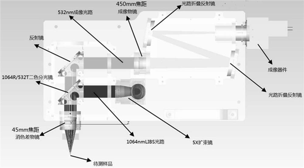
[0044] (1) Plasma excitation device: Nd:YAG laser (Innolas, Germany) was used to generate laser pulses. The laser beam is focused on the sample surface with a small spot size to excite the plasma ( figure 1 ). Specifically, the 1064nm laser is vertically incident on the 5X beam expander from the small hole at the back end of the equipment. After the beam expands and compresses the divergence angle, the laser continues to enter the laser / imaging coaxial system, and is reflected by the 1064R / 532T dichroic beam splitter and turned 90°. Focus on the sample to be tested through the 45mm focal length achromatic objective lens.

[0045] (2) Displacement device: The motorized XYZ stage is triggered by a pulse signal from a digital delayer. As the sample is fixed on...

Embodiment 2

[0049] Example 2: Method for Identifying Tumor Boundary in Lung Cancer Tissue Based on Molecular Spectral Imaging of LIBS Technology

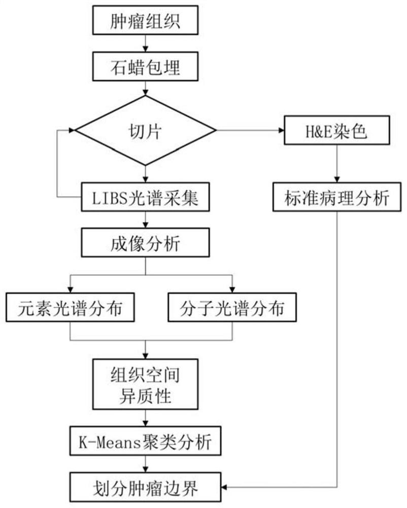
[0050] The method for identifying the tumor boundary of lung cancer tissue based on molecular spectral imaging of LIBS technology in the present invention comprises the following steps:

[0051] 1. Sample preparation

[0052] Samples were collected from lung cancer patients diagnosed as adenophosphocarcinoma after surgical resection, and three types of tissues were collected: cancer tissue, paracancerous tissue, and normal tissue; then, they were paraffin-embedded according to standard pathological methods, and the embedding was performed using a microtome. The paraffin block of the tissue was sectioned until a flat section exposing most of the tissue was obtained, and the subsequent LIBS analysis was performed directly on the sectioned paraffin block.

[0053] At the same time, serial sections of 5 μm were collected for H&E staining and stand...

Embodiment 3

[0065] Example 3: Method for Identifying Tumor Boundary in Lung Cancer Tissue Based on Elemental Imaging of LIBS Technology

[0066] The method for identifying the tumor boundary of lung cancer tissue based on the elemental imaging of LIBS technology of the present invention comprises the following steps:

[0067] 1. Sample preparation

[0068] With embodiment 2.

[0069] 2. Set device parameters

[0070] With embodiment 2.

[0071] 3. Imaging analysis

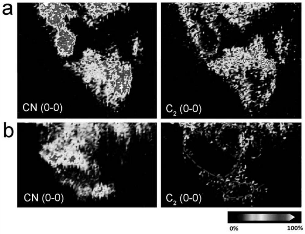
[0072] Perform data preprocessing such as baseline calibration on the collected LIBS spectra, and then extract Na(I) (588.8-589.3nm), Ca(II) (392.9-393.8nm), Mg(I) (279.3-279.8nm), Al (I)(395.9-396.3nm), Si(I)(287.9-288.4nm), Fe(II)(259.8-260nm) and Cu(I)(324.4-324.9nm) peak area, normalized The relative value of the element spectrum peak area of ​​each point in the matrix is ​​obtained. Black corresponds to the relative peak area value of 0%, and white corresponds to the relative peak area value of 100%. The change in t...


PUM


Abstract

The invention provides a method for identifying tumor boundaries by performing molecular spectral imaging on cancer tissues by using a laser-induced breakdown spectroscopy technology, and belongs to the field of imaging. The method comprises the following steps: (1) carrying out molecular spectral imaging on a sample tissue by utilizing a laser-induced breakdown spectroscopy technology, and collecting an LIBS spectrum; (2) after performing baseline calibration on the LIBS spectrum, extracting a CN molecular spectral line or a C2 molecular spectral line to obtain an intensity relative value of the spectral line; (3) drawing a CN molecule or C2 molecule distribution heat map in the sample tissue; (4) carrying out clustering analysis on the CN molecule or C2 molecule distribution heat map to identify the boundary of the tumor region and the necrosis region in the sample tissue. By utilizing the method, the boundary of the tumor area and the necrosis area in the cancer tissue or para-carcinoma tissue of the lung cancer patient can be successfully displayed, and tissue space heterogeneity information is obtained. The method provided by the invention realizes molecular spectral imaging analysis of lung cancer tissues and tumor boundary identification, and has good application prospects and values.

Description

technical field [0001] The invention belongs to the field of imaging, and in particular relates to a method for identifying tumor borders by using laser-induced breakdown spectroscopy to perform molecular spectrum imaging on cancer tissues. Background technique [0002] Chemical elements play an important role in the physiological homeostasis of the human body. Imbalance in the content and distribution of elements may lead to pathological changes, so the content and distribution of elements in biological tissues are helpful for diagnosis and treatment. Studies have shown that element abnormalities may It is related to the occurrence and development of cancer, so people try to analyze the elemental characteristics of cancer tissue, complement common clinical detection methods, and solve difficult problems such as tumor boundary recognition. [0003] In medicine, the color reaction of some special dyes and indicators is usually used to detect metal elements in biological tissu...

Claims


Application Information

Patent Timeline
IPC IPC(8): G01N21/71
CPCG01N21/718
Inventor 林庆宇殷鹏鲲段忆翔
Owner SICHUAN UNIV
Why Eureka
Try Eureka