Preparation method, product and application of antiferromagnetic nanoparticle biological imaging probe

A nanoparticle and antiferromagnetic technology, which is applied in the field of preparation of antiferromagnetic nanoparticle bioimaging probes, can solve the problem of difficulty in controlling the particle size of nanoparticles, the physicochemical properties of magnetization, and the biocompatibility of antiferromagnetic nanoparticles. Poor and difficult in vivo bioimaging and other problems, to achieve good biocompatibility and availability, reduce toxic side effects, and improve the effect of magnetic resonance imaging

Inactive Publication Date: 2021-09-24
ZHEJIANG UNIV
View PDF8 Cites 0 Cited by
  • Summary
  • Abstract
  • Description
  • Claims
  • Application Information

AI Technical Summary

Problems solved by technology

However, the current preparation technology of antiferromagnetic nanoparticles is complicated, and it is difficult to control the physical and chemical properties of nanoparticles such as particle size and magnetization; and the prepared antiferromagnetic nanoparticles have poor biocompatibility and are difficult to be used in living biological imaging.

Method used


Image

  • Preparation method, product and application of antiferromagnetic nanoparticle biological imaging probe
  • Preparation method, product and application of antiferromagnetic nanoparticle biological imaging probe
  • Preparation method, product and application of antiferromagnetic nanoparticle biological imaging probe

Examples


Embodiment 1

[0047] Example 1: Synthesis of oil phase ferromagnetic nanoparticles

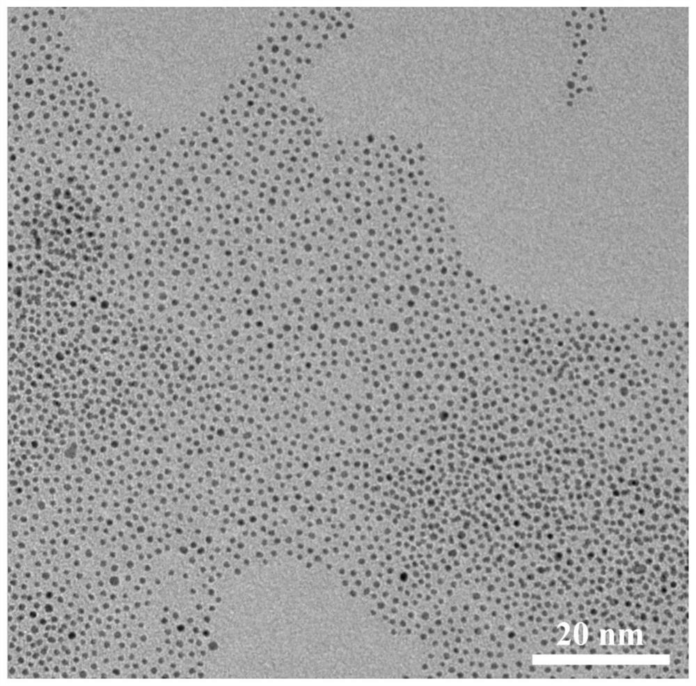
[0048] Dissolve 90 mg of iron acetylacetonate and 100 mg of platinum acetylacetonate in a mixed solution of 415 μl oleylamine, 400 μl oleic acid and 10 ml of dibenzyl ether, react for 30 minutes under an inert atmosphere at 300 ° C, and precipitate with ethanol after the reaction stops to obtain oil phase of antiferromagnetic nanoparticles.

[0049] The morphology of the prepared oil phase ferromagnetic nanoparticles was characterized by transmission electron microscopy, as shown in figure 1 shown.

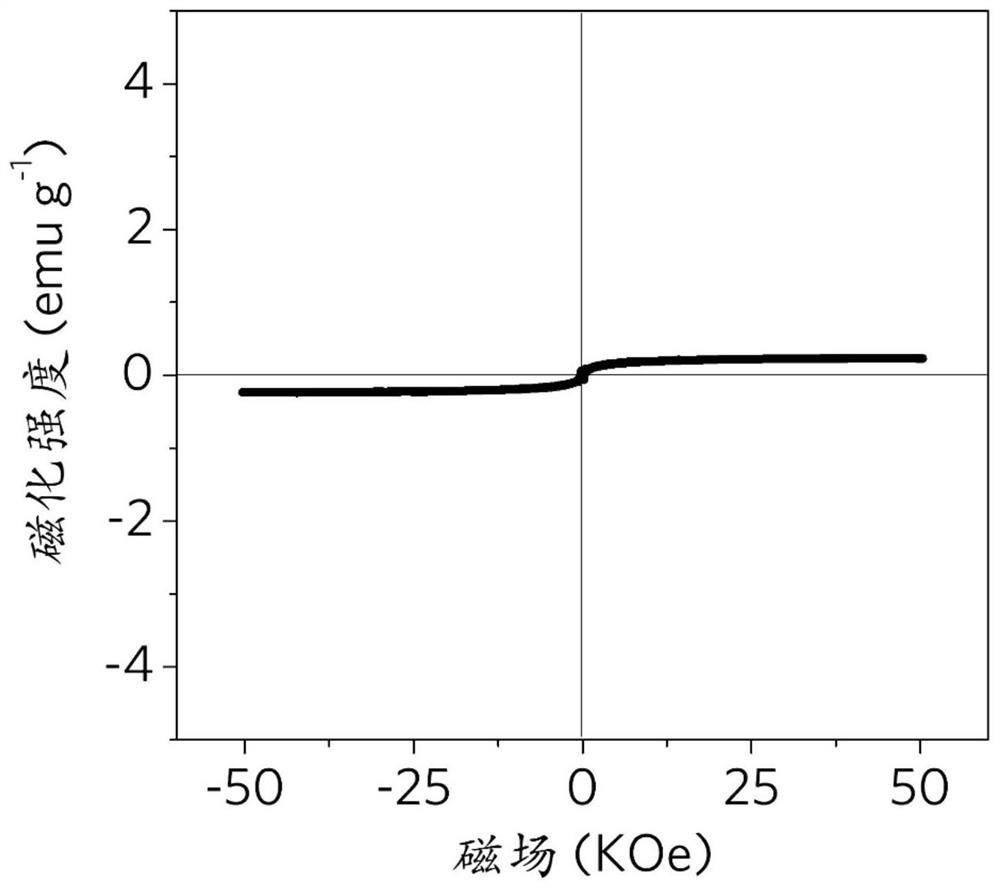
[0050] Using a vibrating sample magnetometer to test its magnetization, the results are as follows figure 2 shown.

[0051] The above results prove that the antiferromagnetic nanoparticles in the oil phase have relatively low magnetization, and the measured magnetization is 0.23 emu / g.

Embodiment 2

[0052] Example 2: Synthesis of oil phase ferromagnetic nanoparticles

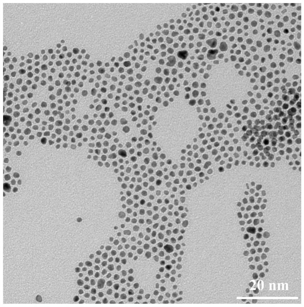
[0053] Synthesized with reference to the preparation process of Example 1, the difference is that the amount of oleylamine and oleic acid added was changed to 800 μl and 830 μl, and antiferromagnetic nanoparticles in the oil phase were also obtained, and the measured magnetization was 4.51 emu / g. Increasing the amount of oleylamine and oleic acid in Example 2 can increase the particle size of the antiferromagnetic nanoparticles in the oil phase.

[0054] The morphology of the oil-phase ferromagnetic nanoparticles prepared by changing the method was characterized by transmission electron microscopy, as shown in image 3 shown.

Embodiment 3

[0055] Example 3: Synthesis of oil-phase ferromagnetic nanoparticles

[0056] Synthesized with reference to the preparation process of Example 1, the difference is that the amount of oleylamine and oleic acid added was changed to 1250 μl and 1200 μl, and antiferromagnetic nanoparticles in the oil phase were also obtained, and the measured magnetization was 4.86 emu / g.

[0057] The morphology of the oil-phase ferromagnetic nanoparticles prepared by changing the method was characterized by transmission electron microscopy, as shown in Figure 4 shown.


PUM


Abstract

The invention discloses a preparation method of an antiferromagnetic nanoparticle biological imaging probe, which comprises the following steps of: dissolving ferric acetylacetonate and platinum acetylacetonate in a mixed solution of oleylamine, oleic acid and dibenzyl ether, reacting for 3-60 minutes under the inert atmosphere condition of 200-300 DEG C, and precipitating a poor solvent to obtain oil-phase antiferromagnetic nanoparticles; performing hydrophilic surface modification on the oil-phase ferromagnetism nanoparticles by using hydrophilic molecules by adopting a ligand exchange method to obtain water-phase antiferromagnetic nanoparticles; and performing a chemical connection reaction on the water-phase antiferromagnetic nanoparticles, and performing targeted surface modification by using a targeted ligand to obtain the antiferromagnetic nanoparticle biological imaging probe. The invention also discloses the antiferromagnetic nanoparticle biological imaging probe prepared by the preparation method and application of the antiferromagnetic nanoparticle biological imaging probe in preparation of a tumour magnetic resonance imaging contrast agent. The antiferromagnetic nanoparticle biological imaging probe has a good high-field magnetic resonance imaging effect, and can be used for performing accurate noninvasive detection on tiny tumours in a living body.

Description

technical field [0001] The invention relates to the field of preparation of magnetic nano-probes, in particular to a preparation method, product and application of an antiferromagnetic nano-particle biological imaging probe. Background technique [0002] Cancer is one of the major diseases that cause human death. Cancer has an insidious onset and rapid progression. Most patients are already in the middle and late stages when they are diagnosed, missing the best opportunity for treatment. If tumors are detected early through accurate diagnosis and intervened before they progress to advanced stages and metastases, the treatment options for cancer patients can be increased and the therapeutic index can be improved. Therefore, early diagnosis of cancer is particularly important. [0003] Magnetic resonance imaging (MRI) technology is widely used in tumor diagnosis and monitoring due to its advantages of non-invasiveness, no radiation and high spatial resolution. This type of ...

Claims


Application Information

Patent Timeline
Patent Type & Authority Applications(China)
IPC IPC(8): A61K49/18A61K49/14
CPCA61K49/1824A61K49/14
Inventor 李方园凌代舜梁泽宇
Owner ZHEJIANG UNIV
Why Eureka
Try Eureka